Download
1 / 72
790 likes | 1.04k Views
Chronic Kidney Disease. Heidi Anderson Erica Bailey Anai Villalobos Katie Pearce . Anatomy of the Kidney. 2 major parts: Cortex Medulla Functional Unit Nephrons Renal Pyramid Renal Pelvis Ureter. Nephrons. 1.) Glomerular Filtration.

E N D
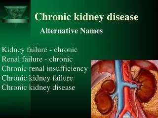
Chronic Kidney Disease Heidi Anderson Erica Bailey Anai Villalobos Katie Pearce
Anatomy of the Kidney • 2 major parts: • Cortex • Medulla • Functional Unit • Nephrons • Renal Pyramid • Renal Pelvis • Ureter
1.) Glomerular Filtration • Fluids and solutes in the blood plasma of the glomerulus pass into the glomerular capsule (glomerular filtrate) • Mechanisms to cause this fluid to be filtered is • High hydrostatic pressure of the blood in the glomerulus • Large number of pores • Substances present in the glomerular filtrate: water, electrolytes, glucose, AA, urea, hormones, and vitamins. • -exclude large molecules that are in the blood.
GFR • The best way to measure levels of kidney function. • GFR= urine volume xinulin conc. In urine inulin conc. In plasma (mg/ml) • Best estimates of GFR: • Inulin clearance • Creatine clearance • Plasma creatinine concentration • Blood urea nitrogen (BUN)
2.) Tubular Reabsorption • Trans-epithelial transport • Most solutes reabsorped completely • Urine volume is regulated by the needs of the body • Active vs. Diffusion • Tm
3.) Tubular Secretion • Movement from the peritubular capillaries into the lumen of the tubule. • Main Substances secreted • H+ • K+ • Some organic anions
Functions of the Kidney • Elimination of wastes, excess water and solutes, and conserves nutrients • Acid-base balance • Renin-angiotensin system • Erythropoietin release • Activation of Vitamin D
Elimination of Waste • Kidney receives 20% of cardiac output, which allows the filtering of approximately 1600 L/day of blood. • This is translated into 1.5 L of urine a day to be excreted, on average • The fluid filtered from the blood plasma is modified and reabsorbed as it travels along the tubules. The remainder finds its way into the ureter, which carries the urine to the bladder from each kidney.
Acid-Base Balance • Kidney is responsible for 2 major activities: • Reabsorption of filtered bicarbonate • Excretion of the fixed acids (acid anion and associated H+) Both of these processes involve secretion of H+ into the lumen by the renal tubule cells Only the 2nd one leads to excretion of H+ from the body. • Acidosis vs. Alkalosis
Renin- Angiotensin System • Regulation Of Blood Pressure
Erythropoietin Release • EPO is a hormone primarily produced by the kidney • Occurs when a drop in blood oxygen level is perceived. • Used to treat anemia. • Glycosylated erythropoietin comes in 3 forms: alpha (the most commonly used type in veterinary medicine), beta (of similar clinical efficacy to alpha) Darbepoetin (which is particularly heavily glycosylated and lasts the longest).
Vitamin D Activation • Parathyroid hormone is able to drive stored calcium and phosphorus from the bones as is vitamin D so these hormones are able to work in concert here but in the kidney they have different functions. In the kidney, while vitamin D saves both calcium and phosphate, parathyroid hormone causes only calcium to be saved and phophate to be dumped. There is a third hormone called “calcitonin” that keeps the blood calcium level from indefinitely rising. When blood calcium starts to get too high, calcitonin is released to begin storing calcium and phosphate back in the bones until it is needed again.
At Risk Populations • Racial Groups: • African Americans • Native Americans • Hispanics • Pacific Islanders • Risk Factors: • Diabetes • Hypertension • Family history of kidney failure
Genetics • The angiotensinogenpromotor G(-6) allele lowers transcription and is inversely associated with hypertension. • the A1166C 3'-UTR variant of angiotensinII type 1 receptor (AT1R) has been associated with CKD. • The AT1R C1166 allele may increase susceptibilitybut only in the presence of hypertension.
What is CKD? • CKD is the gradual loss of the kidney’s ability to filter waste and fluid from the bloodstream. • Nephrons filter waste out of the blood. • Nephrons become damaged and lose their filtering ability overtime. • As more nephrons are damaged, the healthy ones work harder. • Kidneys become scarred or may shrink in size.
Etiology • Diabetes is the number one cause of kidney disease, responsible for about 40% of all kidney failure. • High blood pressure is the second cause, responsible for about 25%. • About 12.2% of Native Americans over the age of 19 have type 2 diabetes. • Diabetes and high blood pressure are the leading causes of CKD.
Etiology-cont. • Other causes include: • Glomerulonephritis (kidney inflammation) • Genetic diseases (i.e. polycystic kidney disease) • Autoimmune diseases (i.e. lupus) • Birth defects • Obstructions caused by problems like kidney stones or tumors
Pathophysiology • As the renal tissue loses function, the remaining tissue increases its performance • ↓ renal function interferes with the ability to maintain fluid and electrolyte homeostasis. • ↓ability to concentrate urine • ↓ability to excrete phosphate, acid and K • ↑creatinine and urea and ↓GFR • Heart failure can result from Na and water overload
Pathophysiology-cont. • ↓production of calcitriol leads to hypocalcemia→osteopenia or osteomalacia. • ↓excretion of phosphate leads to hyperphosphatemia • Secondary hyperparathyrodism is common→renalosteodystrophy. • Normochromic-normocytic anemia (Hct of 20-30%) caused by ↓erythropoietin production due to ↓functional renal mass.
Phosphorus Pathophysiology Cacliphylaxis Decreased renal function Increase blood calcium levels Increased blood phosphorus Decreased active vitamin D Decreased bone mass Increased blood PTH
Cacliphylaxis • Deposition of calcium phosphate in soft tissues • This occurs when the phosphorus-calcium product is too high (greater than 4.5 (mmol/l)2 • Cacliphylaxis is associated with CVD • Calcium phosphate is deposited on heart valves and blood vessels
Diagnosis • Based on laboratory testing of renal function: - Creatinine - GFR - BUN • Urinalysis (check for protein, blood and WBC in urine-which should not be there). • Sometimes renal biopsy. • MRI or ultrasound to check the size.
LABS • Glomerularfiltration rate (GFR) is the best measure of kidney function. • A doctor will order a blood test to measure the serum creatinine level. As kidney function ↓, blood levels of creatinine ↑.
Creatinine • It is a waste product that is passed through the kidneys. • A by-product formed by muscle contractions. It also comes from protein foods we eat. • ↑creatinine may signal that the kidneys aren’t eliminating this waste, leaving it in the body. • Normal range: 0.8-1.4mg/dL
Glomerular Filtration Rate (GFR) • Measures the kidney function and the stage of CKD. • As the kidneys become more damaged, the GFR will decrease. • Normal range: >90, with little or no protein or albumin in the urine.
Blood Urea Nitrogen (BUN) • The BUN test measures the amount of urea in your bloodstream. • Urea is a waste product left over from the protein we eat, which is normally eliminated through the kidneys. • ↑urea mean the kidneys are not getting rid of waste and it remains in the body. • Normal range: 7-20 mg/dL
S/S • CKD is a silent, but devastating disease. • Azotemia • Uremia • ↑↓urination (nocturia) • Fatigue/weakness • Nausea and vomiting • Bruising/bleeding • Uremic frost (crystals in and on skin)
S/S – cont. • Loss of appetite • Edema in feet, ankles, hands, or face • Back pain • Itching • Shortness of breath (fluid can build up I lungs) • Ammonia breath or taste in the mouth
5 Stages of CKD • Help doctors give the best treatment to patients • Each stage requires different tests and treatments
Stage 1 • GFR > 90 ml/min • Kidney damage • Normal or increased function • No symptoms of damaged kidney • People aren’t usually diagnosed unless they are being tested for something else
Stage 2 • GFR 60-89 ml/min • Mild decrease in kidney function • This GFR is normal for some people • People with GFR >60 are still considered to have Chronic Kidney Disease if they have some kind of damage to their kidney
Stage 3 • GFR 30-59 ml/min • Moderate decrease in kidney function • Uremia • Symptoms may start to develop
Stage 4 • GFR 15-29 ml/min • Severe decrease in kidney function • Patients will start thinking about dialysis or transplant • Think about getting a fistula so it has time to mature
Stage 5 • Less than 15 ml/min • Kidney failure with treatment • End stage renal disease • Kidneys are unable to remove waste and fluid from the body
Complications of CKD • Cardiovascular disease • Anemia • High blood pressure • Bone disease
Can CKD be Halted or Reversed? • Cannot be reversed • No known cure (other than kidney transplant) • Can be halted! • Use preventative techniques to halt the progression.
Prevention • Prevent and control high blood pressure • Prevent and control diabetes • Early diagnosis • Routine physical examinations • Stop smoking • Decrease alcohol consumption
Hypertension Prevention/Control • Most important thing to prevent of CKD • Maintain Healthy weight • Exercise to raise your heart rate • Reduce Sodium in diet • Take Medication • Can slow rate of kidney damage by 50%!
Diabetes prevention • Eat a healthy Diet • Fiber, whole grains • Fruits and vegetables • Maintain normal blood glucose levels • Exercise • Obtain and maintain healthy weight
Prevention Programs • National Kidney Foundation’s KEEP • Free screening • Educational materials • Designed to raise awareness • Need a better global effort • Require a lot of man power and funds
Diabetes and Birth • Can lead to complications for the fetus as soon as the first 6-8 weeks of life • CNS deformities • Musculoskeletal deformities • Congenital heart disease • Spontaneous abortion • Large birth weights • Shoulder dystocia
Diabetes and Birth • Large birth weight • If diabetes is not controlled • Baby gets high blood sugar • Baby makes more insulin • Stores the extra calories as fat • “overfed”
Compliance with Diabetes treatments and development of CKD • Study done showing that intensely treated diabetics were 21% less likely to have nephropathy. • Patients who more tightly control their blood sugar are less likely to have renal complications • The longer a patient is noncompliant with diabetes treatments, the greater risk he/she has of developing CKD.