Download
1 / 58
590 likes | 610 Views
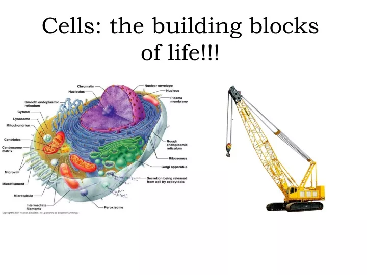
Cells: the building blocks of life!!!. What is a cell?. A cell is the basic structural and functional unit of a living organism What kind of cells can you think of ? . Cells: An Overview Generalized. There are trillions of cells in the human body

E N D
What is a cell? A cell is the basic structural and functional unit of a living organism What kind of cells can you think of ?
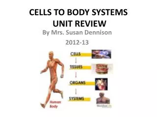
Cells: An Overview Generalized • There are trillions of cells in the human body • Of those trillions, there are over 200 different kinds that vary in size, shape and function
Cells vary in size and shape 1 micrometer (1μm) = 1 x10⁻⁶ m) Red Blood Cell (RBC) 7.5 μm Smooth Muscle Cell 20-500 μm long Human Egg Cell (Ovum) 140 μm Nerve Cell Can be many cm in length White Blood Cell 10-12 μm
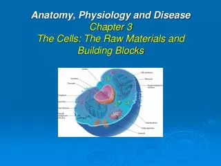
Despite all their differences….ALMOST all human cells have 3 common components • Plasma membrane – the outer layer of the cell • Cytoplasm – fluid inside the cells. Contains organelles • Nucleus – the control center of the cell *****RBC’s do not have a nucleus!!!!
The Plasma Membrane…What is it? • The outer layer of the cell. Think of it as the “traffic controller” for the cell • It is a semi-permeable membrane that is selective about what can enter or leave the cell…the “bouncer” • Separates the body’s 2 main fluid compartments • Intracellular fluid – fluid inside the cell • Extracellular fluid- fluid outside and in between the cells
The Plasma Membrane: Structure Phospholipids 75% Glycolipids 5% Cholesterol 20% Double layer membrane composed primarily of phospholipids
Phospholipids • The polar heads are attracted to water so they lie on the inner and outer surface of the membrane ***remember intracellular fluid and extracellular fluid = water The nonpolar tails avoid the fluid and line up in the center of the membrane
Glycolipids • Lipid with an attached sugar group • Found only in the outer surface of the plasma membrane • Combine with other glycolipids to make glycocalyx (sugar coating)
Glycocalyx • The fuzzy, sticky carbohydrate-rich area surrounding the cell • Every cell has a different pattern of sugars in its glycocalyx; therefore, the glycocalyx provides a very specific biological marker for cell recognition • Essentially I.D. tags for the cell to cell recognition
CholesterolIt’s not the devil! • Wedges between the phospholipid tails • Stabilize the membrane
What 3 factors bind cells together? • Glycoproteins in the glycocalyx act as an adhesive • Wavy contours of membranes fit together in a tongue and groove fashion • Special membrane junctions are formed
Special Membrane Junctions: Tight Junction: • “impermeable” junction helps prevent molecules from passing through the extracellular space between adjacent cells Desmosome: • “anchoring” junctions scattered like rivets along the sides of adjacent cells that prevent separation • Button like plaque on cytoplasmic side held together by thin linker proteins (cadherins) on the cellular side • Thicker protein filaments “lock” together with the plaque on the opposite side to anchor them together • Strong yet flexible junctions
Special Membrane Junctions: Gap Junctions: • Allows chemical substances to pass between adjacent cells • Connected to other cells by a hollow cylinder (connexons) • Ions, sugars, and other small molecules pass through these channels
Cell Junction Junction… What’s Your Function?
Membrane Transport The plasma membrane is a selectively permeable membrane that allows nutrients to enter the cell while keeping unwanted elements out of the cell as well as ridding itself of toxic waste products. Interstitial Fluid (the cellular super highway) • Fluid between the cells that contains nutrients such as vitamins, sugars and amino acids, hormones and neurotransmitters, and waste products
Passive Transport(See Table 3.2 on Page 80) • Simple Diffusion • Facilitated Diffusion • Osmosis • Filtration
Active Transport(See Table 3.2 on Page 80) • Solute Pumping • Vesicular Transport • Exocytosis • Phagocytosis (Endocytosis) • Bulk-phase Endocytosis • Receptor-mediated Endocytosis
Diffusion • The tendency of molecules or ions to scatter evenly throughout the environment • Molecules move from areas of higher concentration to lower concentration
Simple Diffusion • Substances that are nonpolar and lipid soluble (oxygen, carbon dioxide, fat-soluble vitamins, and alcohol) diffuse directly through the lipid bilayer • However, polar and charged particles can selectively pass through channel protein pores if they are small enough Diffusion Model
Facilitated Diffusion Certain molecules (glucose and other simple sugars) are too polar to dissolve in the lipid bilayer and too large to pass through membrane channels so they must be helped across Transport proteins in the plasma membrane allow entrance to the cell bypassing the non polar portion of the cell by engulfing then releasing the molecule into the cell Facilitated Diffusion Model
Osmosis • The diffusion of a solvent, such as water through a selectively permeable membrane • Occurs whenever the water concentration differs on the two sides of the membrane ***Even though water is highly polar, it passes easily through the lipid bilayer**** Osmosis Model
Tonicity • Osmotic imbalances cause cells to shrink or swell until the solute concentration on both sides of the plasma membrane is the same, or the membrane is stretched to it’s breaking point • Tonicity is the ability of a solution to change the tone or shape of cells by altering their internal water volume
Tonicity • Isotonic • solutions with the same concentration of solutes as cells • Cells retain normal shape and have no loss or gain of water • Hypertonic • Solutions with higher concentration of solutes than the cell • Cells in a hypertonic solutions lose water and shrink (crenate) • Hypotonic • Solutions with a lower concentration of solutes than the cell • Cells in a hypotonic solution gain water and swell and sometimes burst (lyse)
Active Transport • Similar to facilitated diffusion in that it needs carrier proteins that combine with the transported substances. • Solute pumps move solutes “uphill” against their concentration gradients
Vesicular Transport • The transport of large particles and macromolecules across the plasma membrane • The substance or cell product to be released is 1st enclosed in a membranous sac called a vesicle • 2 types of vesicular transport • Exocytosis - movement of substances from the cell interior to the extracellular space • Endocytosis – movement of substances from the extracellular space into the cell
Exocytosis • Moves materials out of the cell • Material is carried in a membranous vesicle • Vesicle migrates to plasma membrane • Vesicle combines with plasma membrane • Material is emptied to the outside
Endocytosis • Extracellular substances are engulfed by being enclosed in a membranous vesicle • Types of endocytosis • Phagocytosis—cell eating‖ • Pinocytosis—cell drinking‖ • Receptor mediated Exocytosis/Endocytosis Model
The Cytoplasm • The “stuff” between the plasma membrane and the nucleus • Forms the foundation of the cell and contains the organelles “Little organs”
The Organelles • The “machinery” of the cell • Each organelle “little organ” has a specific job in the cell to maintain the life of the cell
Mitochondria • The “power plants” of a cell providing most of its ATP supply • Carbohydrate, lipid and protein molecules are broken down here and the energy is used to form molecules of ATP • Complex organelles that contain their own DNA, RNA, and ribosomes and are able to reproduce themselves
Ribosomes • Made of protein and RNA, these are the site of protein synthesis (production) • Some are free floating in the cytoplam and others are attached to membranes forming Rough ER
Rough ER • Ribbons of membrane studded with ribosomes, which make all the proteins secreted from the cells • Manufactures the integral proteins and phospholipids that form the plasma membrane –considered a “membrane factory” • Once made, proteins are enclosed in vesicles for transport to the Golgi Aparatus where they are further processed
Golgi Aparatus • Made of stacked, flattened membranous sacs with many tiny vesicles that pinch of for “shipping proteins. • Main function is to modify, concentrate and package the proteins and lipids made by the rough ER. --See figure 3.20 on page 86 • “Packages” are shipped 1 of 3 ways • Vesicle is destined for exocytosis • Vesicle is to become part of the plasma membrane • Vesicles becomes a lysosomes
Lysosomes • Vesicles produced by the Golgi Aparatus that contain digestive enzymes • Function as a cell’s “demolition crew”by • Digesting particles taken in by phagocytosis (esp. bacteria, viruses, and toxins) • Geting rid of worn-out or non-functioning organelles • Performing metabolic functions such as glycogen breakdown and release • Breaking down non-useful tissues such as the webbing between the fingers and toes of a developing fetus • Breaking down bone to release Calcium ions into the blood
Peroxisomes • Membranous sacs containing powerful enzymes (oxidases and catalases) which detoxify harmful substances and neutralize free radicals • Especially numerous in the liver and kidney cells which are very active in detoxification • Free Radicals- normal byproducts of cellular metabolism that can have harmful effects on cells if allowed to accumulate
Check Your Understanding What organelle is the major site of ATP synthesis? What are 3 organelles involved in protein synthesis and how do they interact? How does the function of lysosomes compare to that of peroxisomes?
The Cytoskeleton • The “cell skeleton” – it is a network of rods running through the cystosol • Supports cell structure and aids in cell movement • 3 types of rods from smallest to larges • Microfilaments • Intermediate filaments • microtubules
Microtubules • Cylindrical structures made of tubulin proteins • Support the cell and give it shape • Involved in intracellular and cellular movement • Form the centrioles
Centrioles • Paired cylindrical bodies each composed of 9 triplets of microtubules • Organize a microtubule network during mitosis to form the spindle and asters • Form the bases of cilia and flagella
Cilia • Whip-like cellular extensions on the surface of certain cells Example: cells that line the respiratory tract have cilia that propel mucus laden with bacteria and dust particles upward away from the lungs Flagella • Long tail-like projection formed by centrioles Example: sperm which have one flagellum used for movement NOTE: Cilia propel other substances across the cell’s surface whereas the flagella propels the cell itself
Microvilli “Little Shaggy Hairs” • Tiny finger like extensions of the plasma membrane • Increase the plasma membrane surface are tremendously
The Nucleus • The control center of the cell • Has 3 regions or structures • The nuclear envelope • Nucleoli • chromatin • Most cells only have 1 nucleus but some are multinucleate – having more than 1 nucleus (ex. Skeletal muscles) • All human cells except red blood cells have at least 1 nucleus. RBC’s are the only anucleate cells therefore cannot reproduce and only live for 3-4 months in the blood stream
The Nuclear Envelope • Surrounds the nucleus in a double layer membrane barrier separated by a fluid filled space • The outer membrane is connected with the rough ER of the cytoplasm and studded with ribosomes & pores
Nucleoli • Spherical bodies found within the nucleus • Produce ribosomal RNA molecules for the creation of ribosomes
Chromatin • Uncoiled chromosomes consisting of DNA and histone protein molecules • Histone is responsible for packing long DNA molecules in a compact, orderly way