Download
1 / 47
590 likes | 1.78k Views
CARDIAC OUTPUT. Dr. Grrishma B. At the end of the class the students should know…. Definition of cardiac output (CO), Stroke volume, EDV, ESV, Ejection fraction, Cardiac index and cardiac reserve and their normal values Factors affecting CO Explain the regulation of CO

E N D
CARDIAC OUTPUT Dr. Grrishma B
At the end of the class the students should know… • Definition of cardiac output (CO), Stroke volume, EDV, ESV, Ejection fraction, Cardiac index and cardiac reserve and their normal values • Factors affecting CO • Explain the regulation of CO • List the methods of measurement of CO and learn the principles of common methods AEJ
Definitions and normal values • CARDIAC OUTPUT Amount of blood ejected by each ventricles per minute Normal Value 5-Litres/ min (approximately 8% of the body volume) • STROKE VOLUME volume of blood ejected from each ventricle with each heart beat Normal Value: 70ml Cardiac output = stroke volume x heart rate =70 x 72 = 5040 ml/min (approximately)
CARDIAC INDEX Cardiac output per sq meter of body surface area 5L/1.7m2 =3L/min/m2 Used to standardize the CO in individuals with different body surface areas • END DIASTOLIC VOLUME Volume of blood remaining in each ventricle at the end of diastole is end diastolic volume Normal Value:130 ml
END SYSTOLIC VOLUME Volume of blood remaining in each ventricle at the end of systole is end systolic volume Normal Value:50 ml • EJECTION FRACTION Percentage of end diastolic volume ejected with each beat. EF=SV/EDV Normal Value:65% Index of myocardial performance
CARDIAC RESERVE • Amount of blood that can be pumped by each ventricle in excess of normal cardiac output • Normal Value: 15-25 L/min in non athlete and 20-40L/min in athlete
PHYSIOLOGICAL VARIATIONS • Age • Gender, 10% less in females • Build • High altitude • Exercise • Excitement • Pregnancy • After eating • Posture • Environmental temperature
Pregnancy • Hormones : Adrenal medullary hormone Thyroid hormone etc
Distribution of cardiac output: Cardiac output is distributed to all organs of the body Major part of cardiac output goes to vital organs Brain = 750 ml/min Kidney = 1200 ml/min Heart = 250 ml/min Skin Bones Muscles Liver Intestines Muscles receive large quantities of blood during exercise In the meantime distribution to other parts will be reduced
FACTORS AFFECTING CO • Factors affecting Stroke volume • Factors affecting HR
Factors affecting Stroke volume • Preload- EDV • Frank starling’s law • Venous return - Skeletal pump -Thoracic pump - Abdominal pump - ECF volume - Sympathetic activity
Atrial pump activity -Normal 15-20% -Increased demand (eg. Exercise) • Ventricular compliance -Stretch ability -Cardiomyopathies, pericardial effusion, cardiac tamponade
Myocardial contractility (inotropic effect) • Ventricular mass - Increase in athletes - Decrease in MI • Autonomic activity - Mainly sympathetic stimulation increases the contractility • Hormonal factors - Positive inotropic- catecholamine, Thyroid hormones, Glucagon, Insulin - Negative inotropic- acetylcholine
Chemical factors -Xanthine – inhibit intracellular breakdown of cAMP -Positive inotropic effect -Negative inotropic effect- hypercapnia, hypoxia, acidosis, general anesthetics, toxins • Drugs -Digitalis
Afterload - Peripheral resistance • Vessel Diameter (Vascular hindrance) • Viscosity of blood (Hematological hindrance) • Poiseuille-Hagen formula • Gives the relationship between viscosity of fluid with radius and length of tube • F = πpr4 • 8ήl
F- flow • P-pressure • r-radius • n- viscosity • l- length of the tube • Also Flow= Pressure (p)/ Resistance (R) • Therefore R=8ήl πr4
Viscosity again depends on hematocrit, composition of plasma( plasma proteins), resistance of red cells to deformation, temperature
Factors affecting Heart rate • Mainly autonomic activity • Sympathetic stimulation- increases heart rate • Parasympathetic stimulation- decreases heart rate • Reflex control- Baroreceptor reflex, chemoreceptor reflex,Bainbridge reflex, cushing’ s reflex • Control by higher centers • Hormonal control-thyroxine, catecholamines • Chemical controls
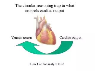
Regulation of Cardiac Output • Intrinsic regulation • Extrinsic regulation
Intrinsic regulation • Autoregulation • Based on preload • Heterometric regulation
Venous return - Skeletal pump -Thoracic pump - Abdominal pump - ECF volume - Sympathetic activity
Atrial pump activity -Normal 15-20% -Increased demand (eg. Exercise) • Ventricular compliance -Stretch ability -Cardiomyopathies, pericardial effusion, cardiac tamponade
Rate induced regulation • Force frequency relation • Increase in frequency increases force of contraction • Increase in number of depolarisation per minute increases Calcium inside the cell • Increase in inner calcium current per depolarisation
Extrinsic regulation • Afterload Homometeric regulation Anrep effect- influence of peripheral resistance on CO
- Peripheral resistance • Vessel Diameter (Vascular hindrance) • Viscosity of blood (Hematological hindrance) • Poiseuille-Hagen formula • Therefore R=8ήl πr4
Viscosity again depends on hematocrit, composition of plasma( plasma proteins), resistance of red cells to deformation, temperature
Neural factors • Sympathetic stimulation- increases heart rate and myocardial contractility • Parasympathetic stimulation- decreases heart rate
Hormonal factors - Positive inotropic- catecholamine, Thyroid hormones, Glucagon, Insulin - Negative inotropic- acetylcholine
Chemical factors Hypercapnia- Directly and indirectly (central and peripheral chemoreceptors) depresses myocardium Hypoxia- moderate to severe hypoxia indirectly (peripheral chemoreceptors) depresses myocardium Acidosis- depresses myocardium by reducing the amount of calcium released from the sarcoplasmic reticulum and decreases the sensitivity of myofilaments to calcium.
Measurement of cardiac output: Direct methods 1. Electromagnetic flow meter (ascending aorta) 2. Cardiometer Indirect methods 1. Fick’s principle 2. Indicator (dye) dilution method 3. Thermo dilution method 3. Echocardiography 4. Esophageal doppler 5. Ballistocardiography 6. X ray method 7. Pulse pressure method
Fick’s principle: This principle suggests that when blood is the only source of a substance, the amount of substance taken up (gained) by the organ or whole bodyis equal to the arterio-venous difference of the substance times the blood flow. Amount of substance taken or given= amount of blood flow per minute x arterio-venous difference Cardiac Output = O2 consumed ml per min/A-V difference.
Hence by knowing the amount of substance removedfrom the blood (concentration in the venousblood) and the previous blood level of substance (arterial concentration), we can calculate the volume of blood flow. To estimate cardiac out put, O2 consumptionor CO2given out can be used.
O2 consumed by the body = 250 ml/min (Spirometry). O2 in arterial blood = 200 ml/ L of blood. O2 in Venous blood =150 ml/ L of blood. CO =250 /50 = 5L/min. O2 content of arterial blood : from any artery. But Venous blood from only Pulmonary artery by cardiac Catheterization from cubital vein. O2 consumption/ min by using a Douglas bag/ spirometer /BMR apparatus (Benedict Roth ) CO2 or Nitrous oxide are also used.
Advantages Result accurate No chemical is injected Disadvantages Catheterization should be done by expert Hospitalization Apprehensive patients increases CO Simultaneous measurement of O2 Difficult in ambulatory patients and in exercise
Hamilton Dye dilution method using Evan’s blue or Indocyanine green: 1ml of Dye injected in to the Basilic vein Serial samples of blood collected from the artery (every 2 sec) Concentration of the dye estimated from samples Photocalorimetrically. Mean concentration determined The volume of plasma in which it was dissolved was calculated Time – concentration curveis plotted to determine average concentration
Time – concentration curve: Concentration Of the dye 0 F = I/ct Time after injection F = Blood flow, I = concentration of the dye injected, c = mean concentration (mean), t = time for first circulation
Advantages lt accurate Disadvantages Injection of dye Cannot be repeated immediately
Thermodilutiondilution technique Similar to dye dilution Cold sterile solution is injected to RA by a catheter through IVC. Resultant temperature change in pulmonary artery is measured. Change in temperature is recorded using thermister Advantages No chemical is injected, saline is harmless Cold dissipates so no recirculation Can be repeated Can be used for children and sick patients Disadvantages Cardiac catheterization
Esophageal doppler transducer Insertion of flexible probe into mid thoracic part of esophagus Doppler transducer is fixed to the tip of the pole which calculates velocity of blood flowing descending aorta. CO from velocity and diameter of the vessel. Echocardiography Non invasive method. Detects direction and velocity of blood in heart and volume of blood flowing through the cardiac valves.
Ballistocardiograhy Vibrations received from each heart beat are received and converted in waveforms by transducer. CO calculated using a special formula. X ray method Radio opaque dye is injected intravenously, size of the heart is detected by serial X rays in systole and diastole. Pulse pressure Method Pulse pressure provides rough idea of CO.
References • Comprehensive Textbook of Medical physiology (Vol 2 first edition) G K Pal • Text book of medical physiology (Vol 2 6 th edition) A K Jain • Essentials of medical physiology (6 th edition) K Sembulingam and PremaSembulingam • Guyton and Hall Textbook of Medical Physiology, 13th Edition AEJ
Reviewed by review committee on 31-12-2018.