Download

1 / 67
680 likes | 843 Views
Movement and the Changing Senses. The communicative link between the human organism and the environment is in part made possible by the senses: vision, proprioception, touch, taste, smell, and hearing. We rely on vision more than any other sense

E N D
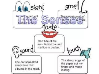
The communicative link between the human organism and the environment is in part made possible by the senses: vision, proprioception, touch, taste, smell, and hearing
We rely on vision more than any other sense • Most movement tasks are initiated by visual information • Nonvisual modalities also influence motor development and performance
Understanding the Mechanics of Vision • Light rays converge and meet at a focal point • The cornea and the fluids in the eye refract (bend) the light rays • The lens can adjust the focal point by changing shape • Relaxation of the ciliary muscles causes the lens to flatten • Contraction of the ciliary muscles causes the lens to become more spherical
Understanding the Mechanics of Vision • Accommodation is the adjustment of the eye to variations in distance • The retina contains two photoreceptors • Rods are responsible for vision in low illumination (night vision) • Cones are responsible for color vision and visual acuity
Understanding the Mechanics of Vision • Macula: an oval yellow spot at the center of the retina • Fovea: (point of best vision) is located here • Cone cells are concentrated • There is an absence of rod cells
Physical Development of the Eye • The eye develops as an outgrowth of the forebrain • Part of the central nervous system • 6 cranial nerves govern vision • At birth • The eye is hyperopic (light focuses behind the retina) • The retina contains mostly rod cells
Physical Development of the Eye • At 1 month postnatal • Cone cells appear • At 8 months postnatal • Macula is mature
Development of Selected Visual Traits and Skilled Motor Performance
Visual Acuity • Degree of detail that can be seen in an object • A Snellen eye chart is used to determine visual acuity
Visual Acuity • This Snellen eye chart is used with children in grades K-1 who may not be capable of letter recognition
Visual Acuity • Static visual acuity • Target and performer are stationary • 20/20 vision means that you see at 20 ft what a person with normal vision sees at 20 ft • 20/100 means that you see at 20 ft what a person with normal vision sees at 100 ft
Visual Acuity • Dynamic visual acuity • Ability to see the detail in moving objects • Ability of the central nervous system to estimate an object’s direction • Ability of the ocular-motor system “to catch” and “to hold” an object’s image on the eye’s fovea long enough to see detail
Visual Acuity and Motor Performance • Both static and dynamic visual acuity play keys roles • Dynamic visual acuity is highly correlated with success in • Field-goal shooting • Ball catching
Visual Acuity and Exercise • Aerobic activities appear to improve visual acuity for up to two hours post-exercise • Increase in acuity due to increase in blood flow and oxygenation to the eye
Age-related eye diseases (ARED) are the leading cause of loss of visual acuity Conditions/diseases Age-related macular degeneration Glaucoma Cataracts Senile miosis Diabetic retinopathy Presbyopia Visual Acuity and Aging
AREDs • Age-related macular degeneration (AMD) • Loss of central vision • Dry form • Breakdown of light sensitive cells in the macula • Not allowed to drive and will have trouble reading • No problem with general movement • Wet form • New blood vessels form behind the retina, leak, and destroy the macula Normal Amsler Grid AMD Visual acuity = 20/50-20/100-total blindness
AREDs • Glaucoma • Leading cause of loss in visual acuity and blindness • Circulating fluids of eye are blocked resulting in high pressure in eye • Loss of peripheral vision • Eventual loss of central vision
AREDs • Cataracts • Clouding of the eye’s lens • Initial symptoms include complaints of glare, colors that seem faded, and increased need for light when reading • Smoking, alcohol, and sun’s UV exposure increase the risk for developing cataracts
AREDs • Senile miosis • Normal loss of light restriction to the eye with age • Decrease in resting diameter of the pupil • Linear decline in the amount of light reaching the retina between 30-60 years
AREDs • Diabetic retinopathy • Complication of diabetes • Vessels in the retina may hemorrhage • Normally clear vitreous humor becomes discolored • Detached retina can occur • Control of blood sugar slows the progression
AREDs • Presbyopia • Inability to focus clearly on near objects as one ages • Bifocal lenses can help “near” vision Normal Prescription Near Vision Prescription
Binocular Vision and Depth Perception • Binocular vision ~ coordinated eye movements • Strabismus ~ misaligned eyes • Common at birth, but diminishes during the first week (moving each eye at random) • Depth perception • A cerebral function based upon information sent by the eye to the brain
Binocular Vision and Depth Perception • The Visual Cliff • Note the mother attempting to coax the infant into crossing the apparent deep (cliff) side • Infants are capable of depth perception • Depth perception is mature at 6 years • Gibson & Walk’s (1960) classic experiment
Depth Perception Held and Hein involved kittens in active and passive movement. The researchers concluded that the active movement benefited the kittens’ development of depth perception. Passive movement did not.
Depth Perception • Motion hypothesis • Individuals must interact with objects that move in order to develop a normal repertoire of visual-spatial skills • Movement does not need to be self-induced
Depth Perception and Sport Success • Accurate depth perception is task specific • Tennis vs. football • Athletes without stereo vision • The central nervous system can use shadows, ball texture, and projectile size to perceive depth
Field of Vision • Refers to the entire extent of the environment that can be seen without a change in fixation of the eye • Normal lateral peripheral vision is a little greater than 90 degrees from straight ahead (180 degrees total) • Normal vertical peripheral vision is 47 degrees above and 65 degrees below visual midline
Field of Vision • Infant’s field of vision • At 2 months ~ peripheral vision is 15 degrees lateral of central vision • At 7 weeks ~ peripheral vision is 35 degrees lateral of central vision
Field of Vision • David’s 1987 experiment examining peripheral vision processing during the performance of a catching task
Field of Vision • David’s experiment • Assessed peripheral vision in children and adults in real-world settings • Peripheral information given to subjects during a performance of ball-catching • Required dual processing of information • 9 year olds made significantly more mistakes in ball catching compared to a single task performance
Aging and Depth Perception and Field of Vision • Both disease (AMD) and anatomical facial changes may cause a loss of depth perception/field of vision with age • Change in facial structure • Senile ptosis • Drooping of the eyelid • Loss of fat tissue around orbital socket
Eye Dominance • Refers to the ability of one eye to lead the other in tasks involving visual tracking and visual fixation • Hole-in-card test • Established between ages 3-5 years
Eye Dominance • Unilateral dominance • Right-eyed and right-handed • Left-eyed and left-handed • Crossed-lateral dominance • Right-eyed and left-handed • Left-eyed and right handed
Eye Dominance • Eye dominance and motor performance • Unilaterals are superior to crossed-laterals in several motor tasks • However, crossed-laterals may have an advantage in baseball batting • A right handed batter with left-eye dominance • Dominant eye is closer to the pitcher • This trait is more common among baseball players than in the general population
Tracking and Object Interception • Tracking an object allows the performer to gain important information about the flight path of the object • Smooth pursuit system • Matching of eye movement speed and speed of a projectile • Saccadic eye-movement system • Corrects differences between projectile location and eye fixation
Tracking and Object Interception • Bassin anticipation time • Coincidence-anticipation • Process involving object interception
Factors influencing success in tracking and interception Object speed Object predictability Viewing time Gender Age Sport participation and video games may be a better predictor than age Boys perform better than girls Very slow and very fast moving objects result in greater performance error Tracking and Object Interception
Blindness • A definition of blindness is based upon distance vision • Ranges from 20/200 (80% loss of vision) to total blindness • Because visual curiosity elicits movement, the unsighted child is not visually motivated to explore the unseen world
Head and trunk control Curiosity encourages lifting head and trunk in sighted children Unsighted child fusses when in a prone position – parent places child on the back which does not help with head and trunk control Independent sitting Occurs in sighted children between 4 and 8 months An unsighted child can perform this task between 4 and 8 months if the parents have prepared the child Need control over head, neck, and trunk Blindness
Creeping By 10 months a sighted child can support him/herself on hands and knees to creep and explore An unsighted child has no enticement to explore Noise-making toys help the unsighted child to creep Independent walking Both sighted and unsighted children are able to walk independently at the same time However, this task is usually delayed in unsighted children Blindness