Download
1 / 35
460 likes | 935 Views
Biological Bases of Behavior. 7: Audition, the Body Senses, and the Chemical Senses. Sound Waves. 7. 2. Divisions of the Ear . Outer ear: Channel to tympanic membrane Middle ear: Ossicles Inner ear: Cochlea. 7. 3. The Cochlea . The cochlea is formed from three chambers:

E N D
Biological Bases of Behavior 7: Audition, the Body Senses, and the Chemical Senses
Sound Waves 7.2
Divisions of the Ear • Outer ear: • Channel to tympanic membrane • Middle ear: • Ossicles • Inner ear: • Cochlea 7.3
The Cochlea • The cochlea is formed from three chambers: • Scala vestibuli and scala media are separated by a membrane • Scala tympani and scala media are separated by the basilar membrane • Hair cells within the organ of Corti transduce sound waves into nerve impulses • The organ of Corti consists of • Basilar membrane (the base) • Tectorial membrane (the roof) • Hair cells in between 7.4
Auditory Hair Cells • Two types of hair cells are located within the human organ of Corti • Inner hair cells (approximately 3500) form a single line of cells along the basilar membrane • Destruction of inner hair cells eliminates hearing • Outer hair cells (approximately 12,000) are arranged in three rows along the basilar membrane • Outer hair cells serve a structural function • Cilia project from the top of each hair cell • The tectorial membrane is attached to the outer hair cell cilia • When sound waves move the basilar and tectorial membranes, the cilia bend in one direction or the other • Shear of the cilia generates a receptor potential that releases a neurotransmitter 7.5
Cilia tips are joined by a fiber link Cilia movement produces tension of the link which opens an ion channel in the adjacent tip Calcium and potassium ions flow into the cilia and produce a depolarization Auditory Transduction 7.6
Auditory Pathways • Afferent pathways: • Spiral (cochlear nerve) ganglion • Through cochlear nuclei • To superior olivary complex • To inferior colliculus • To medial geniculate • To auditory cortex • Efferent pathway: • Olivocochlear bundl • From superior olivary complex 7.7
Place Coding of Pitch • Different frequencies produce maximal distortion of basilar membrane • Sound vibration produces a traveling wave • High frequency: near base of basilar membrane • Moderate frequency: near apex of basilar membrane • Different regions of the basilar membrane project to different areas of auditory cortex • base to medial and posterior, apex to lateral and anterior • Throughout the auditory system there is a tonotopicrepresentation in which adjacent neurons receive signals from adjacent areas of the basilar membrane • Place coding can account for medium to high sound frequencies, low frequency sounds are coded by rate of firing 7.8
Support for Place Theory • Observations of traveling waves by von Bekesy • Different frequencies produce maximal displacement at different points along the basilar membrane • Antibiotics • Induce hair cell loss first at base of basilar membrane, which produces a loss of hearing for high frequency sounds • Cochlear implants restore speech perception by stimulating different regions of the basilar membrane 7.9
Perception of Spatial Location • Arrival time difference for high frequency sound 7.10
Perception of Spatial Location • Horizontal locations • Arrival time difference for high frequency sound • Arrival phase difference for low frequency sound • Intensity difference for high frequency sound • Vertical location • All above methods won’t work for vertical middle-plane • Timbre difference pinna (external ear) • Same for front versus back 7.11
Analysis of the Auditory System • The various components of the auditory system detect sounds, determine sound location, and recognize sound identity • Ventral (what) system: auditory cortex, inferior frontal gyrus • Dorsal (where) system: superior parietal cortex, superior frontal gyrus • Lesions placed at different levels of the auditory system: • Auditory association cortex: auditory agnosia • Bilateral auditory cortex: animal can detect pitch, intensity diff, but not “tunes” • Lateral lemniscus: animal is deaf 7.12
Anatomy of the Vestibular Apparatus • Vestibular sac: • One of a set of two receptor organs (utricle and saccule) in each inner ear that detects changes in the tilt of the head, related to nausea • Semicircular canal: • One of the three ring-like structures of the vestibular apparatus that detect changes in head rotation, related to dizziness and nystagmus (rhythmic eye movements) • Ampulla: • An enlargement in a semicircular canal; contains the cupula. • Cupula: • A gelantinous mass found in the ampulla of the semicircular canals; moves in response to the flow of the fluid in the canals. 7.13
The Vestibular Pathway • The receptor cells: • Similar to the hair cells found in the cochlea; method of transduction is also similar to hair cells of the cochlea. • Vestibular ganglion: • A nodule on the vestibular nerve that contains the cell bodies of the bipolar neurons that convey vestibular information to the brain. 7.14
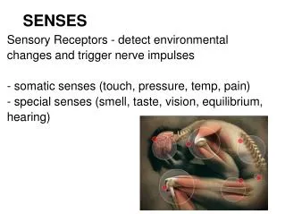
Somatosenses • The somatosenses provide information relating to events on the skin and to events occurring within the body • Cutaneous senses receive various signals from the skin that form the sense of touch • Pressure • Vibration • Heating/cooling • Stimuli that damage tissue (and produce pain) • Kinesthesia provides information about the body position and movement • Kinesthetic signals arise from receptors located within the joints, tendons, and muscles • Organic senses arise from receptors in and around internal organs 7.16
Morphology of Skin Epidermis Dermis 7.17
Cutaneous Senses • Three different sensations are reported to the brain by receptors localized within skin • Touch involves perception of pressure and vibration of an object on the skin • Pacinian corpuscles detect rapid vibrations, with large receptive fields • Meissner’s corpuscle (glabrous skin only) detect slow vibrations • Ruffini corpuscles (plus Merkel’s disks on glabrous skin) respond to indentation, with slow adaptation • Free nerve endings around hairs detect movements of hairs • Temperature is detected by warmth and cold receptors • Receptor activation is relative to the baseline temperature • The receptors (free nerve endings) lie at different levels of the skin (cold are close to the surface of the skin) • Pain is associated with skin tissue damage • Detected by free nerve endings (nociceptors) 7.18
The dorsal columns carry information related to touch (precisely localized) The spinothalamic tract carries pain and temperature signals (poorly localized) Mostly contra lateral Somatosensory Pathways 7.19
Somatosensory cortex is organized into columns There may be 5-10 cortical maps of the body surface Somatosensory Pathways 7.20
Pain • Pain serves a functional role for survival • Persons lacking pain receptors are at great risk • Pain stimuli induce species-typical escape and withdrawal responses • Pain is a motivational force that can activate behavior • Pain involves tissue destruction induced by • Thermal stimuli • Mechanical force • Pain reception is poorly localized (as is temperature) • Pain involves an emotional component (that can be used to modify the magnitude of pain perception) 7.21
Pain Receptors • Receptors for pain (nociceptors) • Free nerve endings networks within the skin that respond to intense pressure • Free nerve endings that respond to heat, acids, and capsaicin (the active ingredient in chili peppers) • Receptors that are sensitive to ATP • Pain receptors are found in: • Skin • Sheath around muscles, internal organs • Cornea of the eye • Pulp of the teeth • Pain receptors are activated by mechanical, chemical stimulation 7.22
Analgesia • Analgesia refers to the reduction of the perception of pain • Analgesia can be induced by external and internal stimuli • Hypnosis • Massage • Acupuncture • Opiates • Placebo • Attention shifts • Pain stimuli activate primary somatosensory cortex (sensory) and anterior cingulate cortex (emotional) • anterior cingulate cortex is involved in the aversiveness of pain • hypnosis and PET scanner study: intensity/unpleasantness 7.23
Opiates and Pain • Exogenous opiates (drugs such as opium, morphine, and heroin) induce analgesia • Environmental stimuli which can activate endogenous opioids also induce analgesia • Certain analgesia could be blocked by Naloxon • Naloxone reversibility is taken as an indication of opiate involvement (such as acupuncture induced analgesia) • Focal brain stimulation can reduce pain • At periaqueductal gray matter in particular is effective • Brain stimulation activates a descending pathway that modulates pain (Basbaum and Fields model) 7.24
Opiate-Induced Analgesia Circuit • Why have pain sensors and analgesia circuit? (experiments on rats,Maier etal 1982) 7.25
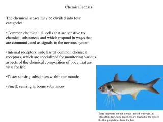
Gustation • Gustation is related to eating foods and drinking liquids • Molecules within the food dissolve in saliva and activate one of four receptor types • Each receptor type provides information about a food • Sweet: safe foods • Salty: source of sodium ions • Bitter: poisonous foods • Sour: spoiled foods • Flavor involves a mixture of taste and olfaction 7.26
Transduction of Taste • Taste molecules bind with a receptor, alter membrane potential, and induce receptor potentials • Saltiness: best stimulus is sodium chloride • Receptor for saltiness may be a simple sodium channel • Sourness receptors respond to hydrogen ions present in acid solutions • Bitterness: typical stimulus is an alkaloid (e.g. quinine) • Receptors involve a hydrophobic residue • Sweetness: typical stimulus is a sugar • Receptors have a hydrogen ion site 7.27
Gustatory Processing • Gustatory information is transmitted through cranial nerves 7 (anterior tongue), 9 (posterior tongue), and 10 (palate and epiglottis) • First relay station for taste information is the nucleus of the solitary tract (medulla) • Taste information is then transmitted to primary gustatory cortex, to the amygdala, and to the hypothalamus • Recordings from chorda tympani (7th cranial nerve) indicate that taste fibers respond to more than one taste quality and to temperature • In cortex, the major groups of taste-sensitive neurons were salty and sweet 7.30
Olfaction • Olfaction is the second chemical sense • The stimulus for odor (known as ordorants) consists of volatile substances having a molecular weight in the range of approximately 15 to 300 • Almost all odorous compounds are lipid soluble and of organic origin • For humans, olfaction is the most enigmatic of the modalities • Flavor involves a mixture of taste and olfaction 7.32
Anatomy of the Olfactory Apparatus • Olfactory epithelium: • The epithelial tissue of the nasal sinus that covers the cribiform plate; contains the cilia of the olfactory receptors. • Olfactory bulb: • The protrusion at the end of the olfactory tract; receives information from the olfactory receptors. • Mitral cell: • A neuron located in the olfactory bulb that receives information from olfactory receptors; axons of mitral cells bring information to the rest of the brain. • Olfactory glomerculus: • A bundle of dendrites of mitral cells and associated terminal buttons of the axons of olfactory receptors. 7.33