Download
1 / 24
280 likes | 676 Views
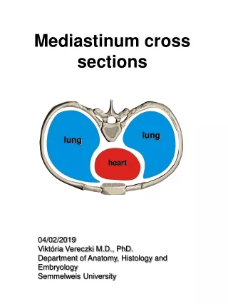
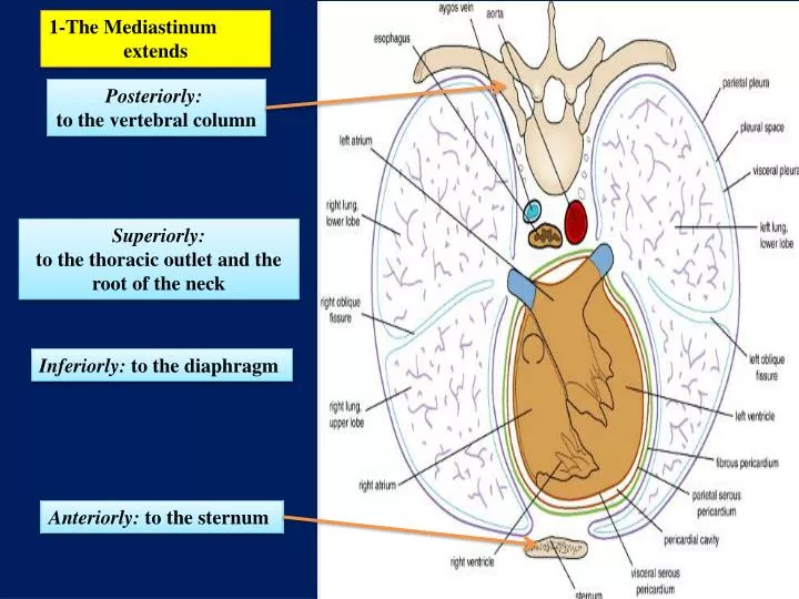
1-The Mediastinum extends . Posteriorly: to the vertebral column. Superiorly: to the thoracic outlet and the root of the neck . Inferiorly: to the diaphragm. Anteriorly: to the sternum. An imaginary plane passing from the sternal angle anteriorly to

E N D
1-The Mediastinum extends Posteriorly: to the vertebral column Superiorly: to the thoracic outlet and the root of the neck Inferiorly: to the diaphragm Anteriorly: to the sternum
An imaginary plane passing from the sternal angle anteriorly to the lower border of the body of the fourth thoracic vertebra posteriorly divides the mediastinum into: SUPERIOR AND INFERIOR MEDIASTINA Which one is larger?
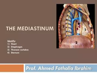
THE INFERIOR MEDIASTINUM is further subdivided into: 1-THE MIDDLE MEDIASTINUM consists of the pericardium and heart 2-THE ANTERIOR MEDIASTINUM is a space between the pericardium and the sternum 3-THE POSTERIOR MEDIASTINUM lies between THE PERICARDIUM And THE VERTEBRAL COLUMN
is bounded in front by the manubrium sterni and behind by the first four thoracic vertebrae THE SUPERIOR MEDIASTINUM contains From anterior to posterior posterior THYMUS LARGE VEINS? LARGE ARTERIES? TRACHEA ESOPHAGUS THORACIC DUCT SYMPATHETIC TRUNKS anterior
Main contents! THE SUPERIOR MEDIASTINUM THYMUS LARGE VEINS, right and left brachiocephalic veins and the upper half of superior vena cava. LARGE ARTERIES, arch of the aorta with its three large branches. TRACHEA ESOPHAGUS THORACIC DUCT (g) phrenic nerves, vagus nerves and SYMPATHETIC TRUNKS Anterior Posterior Cross-section through the superior mediastinum at the level of vertebra TIII.
THYMUS • Involved in the early development of the immune system, • the thymus is a large structure in the child, • begins to atrophy after puberty • In the elderly adult, it is barely identifiable as an organ, consisting mostly of fatty tissue
the right internal jugular veins Large Veins of the Thorax the right subclavian 1-Brachiocephalic Veins A-The right brachiocephalic vein: formed by the union of the right subclavian and the right internal jugular veins (note: it is shorter and vertical)
Is formed by the union of the LEFT subclavian and the LEFT internal jugular veins ( note: It passes obliquely and it is longer) It joins the right brachiocephalic vein to form the superior vena cava B-The left brachiocephalic vein:
Superior Vena Cava • The superior vena cava contains all the venous blood from the head and neck and both upper limbs • is formed by the union of the two brachiocephalic veins • It passes downward to end in the right atrium of the heart • The vena azygos joins the superior vena cava just before it enters the pericardium
Azygos Veins The azygos veins consist of: c-THE SUPERIOR HEMIAZYGOS VEIN a-THE MAIN AZYGOS VEIN b-THE INFERIOR HEMIAZYGOS VEIN
Inferior Vena Cava The inferior vena cava pierces the central tendon of the diaphragm opposite the eighth thoracic vertebra and almost immediately enters the lowest part of the right atrium
Large Arteries of the Thorax Aorta The aorta is the main arterial trunk that delivers oxygenated blood from the left ventricle of the heart to the tissues of the body. It is divided for purposes of description into the following parts: A-ASCENDING AORTA B-ARCH OF THE AORTA C-DESCENDING: THORACIC AORTA (above diaphragm) ABDOMINAL AORTA (below diaphragm)
A-Ascending Aorta • The ascending aorta lies within the fibrous pericardium • (what does this mean?) • Begins at the base of the left ventricle • Ends at the level of the sternal angle, where it becomes continuous with the arch of the aorta • At its root it possesses three bulges, the sinuses of the aorta Branches The right coronary artery The left coronary artery
B-Arch of the Aorta • is a continuation of the ascending aorta • (what does this mean?) • Ends at the level of the sternal angle where it becomes continuous with the descending aorta. • Branches • a-THE BRACHIOCEPHALIC ARTERY • arises from the convex surface of the aortic arch • It divides into: • 1-THE RIGHT SUBCLAVIAN ARTERY • 2-RIGHT COMMON CAROTID ARTERY
b-The left common carotid artery • Arises from the convex surface of the aortic arch • enters the neck behind the left sternoclavicular joint. c-The left subclavian artery • Why we call it subclavian? • arises from the aortic arch • Runs in a groove in the first rib
C- Descending Thoracic Aorta • lies in the posterior mediastinum • begins as a continuation of the arch of the aorta • (opposite the sternal angle). • At the level of • the 12th thoracic vertebra, • it passes behind the diaphragm (through the aortic opening) in the midline and becomes continuous with the abdominal aorta. Branches READ ONLY 1-Posterior intercostal arteries are given off to the lower nine intercostal spaces 2-Pericardial, esophageal, and bronchial arteries are small branches that are distributed to these organs.
What do you think about the aorta and mediastina?
Thoracic Duct • The thoracic duct begins below in the abdomen as a dilated sac, • THE CISTERNA CHYLI • It ascends through the aortic opening in the diaphragm • enters the beginning of • the left brachiocephalic vein. Passes through superior and posterior mediastinum Read only The thoracic duct thus conveys to the blood all lymph from the lower limbs, pelvic cavity, abdominal cavity, left side of the thorax, and left side of the head, neck, and left arm
Nerves of the Thorax • Vagus Nerves • Cranial nerve 10 • The right vagus nerve • The left vagus nerve • Located in • the neck • Thorax: passes through both • superior and posterior mediastinum
Phrenic Nerves • The phrenic nerves arise from the anterior rami of the third, fourth, and fifth cervical nerves • It passes in front of the root of the lungs Read only Its terminal branches pass through the caval opening in the diaphragm to supply the central part of the peritoneum on its underaspect.
Thoracic Part of the Sympathetic Trunk • The thoracic part of the sympathetic trunk is continuous above with the cervical and below with the lumbar parts of the sympathetic trunk. • : passes through both • superior and posterior mediastinum Read only The first ganglion is often fused with the inferior cervical ganglion to form the stellate ganglion
The esophagus has three anatomic and physiologic constrictions Esophagus • is a tubular structure about 10 in. (25 cm) long • is continuous above with the laryngeal part of the pharynx opposite • the sixth cervical vertebra. • It passes through the diaphragm at the level of the • 10th thoracic vertebra • to join the stomach where the pharynx joins the upper end 1 2 where the aortic arch and the left bronchus cross its anterior surface These constrictions are Read only 3 where the esophagus passes through the diaphragm into the stomach • passes through both • superior and posterior mediastinum