Download
1 / 36
380 likes | 777 Views
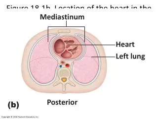
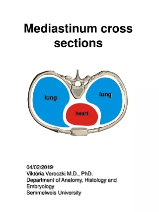
Mediastinum and Heart. Sanjaya Adikari Department of Anatomy. Mediastinum. Mediatinum. Mediastinum is the space between the two lungs It is bounded superiorly by the superior thoracic aperture inferiorly by diaphragm anteriorly by the manubrium and sternum

E N D
Mediastinum and Heart Sanjaya Adikari Department of Anatomy
Mediatinum • Mediastinum is the space between the two lungs • It is bounded • superiorly by the superior thoracic aperture • inferiorly by diaphragm • anteriorly by the manubrium and sternum • posteriorly by the vertebral column • laterally by the two lungs
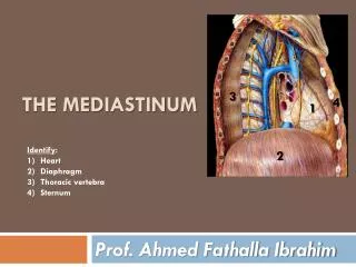
Divisions of the mediastinum Name the structures present at the plane passing through sternal angle
Superior mediastinum Inferior mediastinum Anterior mediastinum Middle mediastinum Posterior mediastinum
Superior mediastinum • What are the boundaries? • ………………………………………….. • ………………………………………….. • ………………………………………….. • What are the contents? • ………………………………………….. • ………………………………………….. • …………………………………………..
Anterior mediastinum • What are the boundaries? • ………………………………………….. • ………………………………………….. • ………………………………………….. • What are the contents? • ………………………………………….. • ………………………………………….. • …………………………………………..
Middle mediastinum • What are the boundaries? • ………………………………………….. • ………………………………………….. • ………………………………………….. • What are the contents? • ………………………………………….. • ………………………………………….. • …………………………………………..
Posterior mediastinum • What are the boundaries? • ………………………………………….. • ………………………………………….. • ………………………………………….. • What are the contents? • ………………………………………….. • ………………………………………….. • …………………………………………..
Heart • Covered by the pericardium • Pericardium has two layers • Fibrous pericardium • Serous pericardium • Visceral layer covering the heart surface • Parietal layer lining the fibrous pericardium
Heart… • Define the different borders and surfaces of the heart • Name the chambers contributing to form the surfaces and borders • Describe the blood supply of the heart
Left coronary artery Right coronary artery
Right coronary artery (RCA) • Arises from right aortic sinus • Gives off SA nodal, right marginal, AV nodal branch and posterior interventricular branch • Dominance is decided by which of the two main coronary arteries gives rise the posterior interventricular branch • Right dominance 67 %, so most commonly the diaphragmatic surface is supplied by RCA
Area supplied by RCA • Right atrium • Most of right ventricle • Diaphragmatic surface of left ventricle • Posterior 3rd of interventricular septum • SA node in 60% • AV node in 80%
Left coronary artery (LCA) • Arises from left aortic sinus • Divides into two branches early; anterior interventricular and circumflex branches • Left dominance 33 % • Left marginal artery is a branch of the circumflex artery
Area supplied by LCA • Left atrium • Most of left ventricle • Anterior wall of RV adjacent to the LV • Anterior two thirds of interventricular septum • AV bundle • SA node in 40% • AV node in 20%
Coronary anastomoses • Coronary artery branches are end arteries • There are anastomoses between branches • Between posterior and anterior interventricular branches • Between RCA and circumflex artery • Between right and left conus arteries
Coronary anastomoses… cont. • There are coronary – extracoronary anastomoses at arteriole level. Mostly pericardial vessels are involved. • Anastomoses in the interventricular septum and within the posterior wall of the left ventricle are more important than the surface anastomoses
Blood supply of the heart wall • Intramuscular, penetrating arteries arising from the coronary arteries connect them to a subendocardial plexus of arteries • During systole, due to contractile forces, the blood flow to this plexus stops almost completely
Cardiac veins… cont. • Main cardiac veins include, • Great, middle and small cardiac veins • Oblique vein draining left atrium • Posterior vein of the left ventricle • Anterior cardiac veins • The anterior cardiac veins drain directly into the right atrium • Other veins drain into the coronary sinus • Coronary sinus drains into the right atrium • Thebesian veins or venae cordis minimae open directly into the respective chambers of the heart
extracoronary arteries arterioles thebesian veins any chamber Summary of coronary circulation coronary arteries arterioles capillaries veins anterior cardiac veins (great/middle/small cardiac veins) right atrium coronary sinus
Myocardial Infarction (MI) • Myocardial death due to sudden loss of blood supply. • Result of coronary artery disease and thromboembolsim • Common sites of coronary artery occlusion, • AIV branch of LCA (40-50%) • RCA (30-40%) • Circumflex branch of LCA (15-20%)