Download

1 / 65
850 likes | 2.04k Views
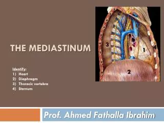
Mediastinum. Dr.Hassan Shaibah. Chest Cavity. Mediastinum. pleurae & lungs. pleurae & lungs. Mediastinum. Mediastinum. Extends superiorly to the thoracic outlet , root of the neck & inferiorly to the diaphragm .

E N D
Mediastinum Dr.HassanShaibah
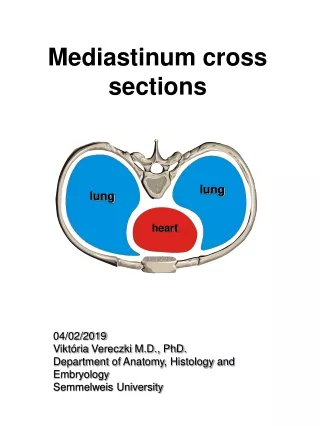
Chest Cavity Mediastinum pleurae & lungs pleurae & lungs
Mediastinum • Extends superiorly to the thoracic outlet ,root of the neck &inferiorlyto the diaphragm. • extends anteriorly to the sternum & posteriorly to the vertebral column. It contains : • thymus, trachea, thoracic duct ,the heart esophagus, large blood vessels, lymph nodes, vagus & phrenic nerves, & sympathetic trunks.
trachea phrenic& vagus nerves large blood vessels thymus Heart Some Contents Mediastinum
esophagus trachea large blood vessels phrenic& vagus nerves Some Contents Mediastinum
The mediastinum is divided by an imaginary plane passing from sternal angle to the Intervertebral discsT4 & T5 into: • superior mediastinum • inferior mediastinum
MediastinumDivsion sternal angle superior 4 5 inferior
The inferior mediastinum subdivided into: • anterior mediastinum, a space between the pericardium and the sternum • Middle mediastinum pericardium and heart • posterior mediastinum, between “pericardium &vertebral column”
* Anterior MIDDLE Posterior *inferior mediastinumm superior
Superior Mediastinum is bounded: • frontby manubriumsterni • behind by first 4 thoracic vertebrae. It contains: • (a) Thymus, (b) large veins, (c) large arteries, (d) trachea, (e) esophagus and thoracic duct, (f) nerves
1 manubrium 4
nerves RT & LT brachiocephalic v. LT common carotid a. Brachiocephalic Trunk thymus
Inferior MediastinumBondries front Body of sternum 5 behind lower 8 thoracic vertebrae 12
Inferior Mediastinum It contains: (a) Thymus, (b) heart within the pericardium. (c) phrenic nerves (d) esophagus and thoracic duct, (e) descending aorta (f)Azygous venous system (g) sympathetic trunks
1)Thymus 3)esophagus 2)heart within the pericardium
Anterior mediastinum, a space between the pericardium and the sternum Middle mediastinumpericardium and heart will be discussed with Cardiovascular block
Posterior Mediastinum Boundaries: . Sup. transverse thoracic plane 5 Ant. pericardium Post. bodies of the vertebral column 12 Inf.diaphragm laterally the pleura (on either side)
Contents 1)Descending aorta
2)Azygos venous system 3)Thoracic duct
9)vagus nerve Vagal plexus
Contents • artery • descending thoracic aorta • Veins • azygos vein • the sup. & inf. hemiazygos vein • nerves • vagus nerve • Sympathetic trunks • esophagus • thoracic duct
Azygos Venous system: • consist of: 2)superior hemiazygos v. • azygos v. • 3)Inferior hemiazygos v.
They drain blood from: • posterior intercostal spaces • posterior abdominal wall • pericardium • diaphragm • bronchi • esophagus.
Azygos Veinformed by union of: SVC T5 Azygos V. 1)right subcostal v. 2)right ascending lumbar v. IVC
Tributaries: Superior hemiazygos veins. RT superior intercostal v. 8 lower right posterior intercostal v. Inferior hemiazygos veins Mediastinal veins.
1)Azygos Vein formed by union of the right ascending lumbar vein and the right subcostal vein. b. It ascends through aortic opening in the diaphragm on the right side of the aorta to the level of the fifth thoracic vertebra. • Arch over the root of the right lung to empty into the SVC e. The azygos vein tributaries are: a. The 8 lower right posterior intercostal veins. b. The right superior intercostal vein. c. The superior and inferior hemiazygos veins. d. Mediastinal veins.
Inferior Hemiazygos Vein • It is formed by the union of the left ascending lumbar vein & left subcostal vein. • It ascends through the left crus of the diaphragm at T8. • turns to the right and joins the azygos vein. • It receives as tributaries some lower left intercostal veins and mediastinal veins.
Superior Hemiazygos Vein • It is formed by the union of the 4 to the 8 intercostal veins. • It joins the azygos vein at the level of the T7.
Mediastinum lymph • Lymph nodes draining the lungs, mediastinalstructures empty into the : bronchomediastinal trunks & thoracic duct.
left Brachiocephalic v. Thoracic Duct begins in the abdomen as a dilated sac (cisternachyli) • Asend to the root of the neck to empty into beginning of the left Brachiocephalic vein cisternachyli
1)left jugular trunk 2)LT Subclavian trunk At the root of the neck, the thoracic duct receives: . 3) broncho-mediastinal lymph trunks.
The thoracic duct carries lymph from: • i. The lower limbs. • ii. The pelvic cavity. • iii. The abdominal cavity. • vi. The left side of the thorax. • v. The left side of the head, neck. • vi. The left arm.
1)RT jugular 2)RT subclavian Right Lymphatic Duct formed by: It opens intobeginning Right brachiocephalic vein. 3)bronchomediastinal trunks
Thoracic Part of the Sympathetic Trunk 1. continuous above with the cervical and below with the lumbar parts of the sympathetic trunk. 2. It is the most laterally placed structure in the mediastinum. 3. It runs downward on the heads of the ribs. 4. It leaves the thorax on the side of the body of the T12 by passing behind the medial arcuate ligament.
5.. The sympathetic trunk has 12 (often only 11) segmentally arranged ganglia, each with white and gray ramuscommunicans passing to the corresponding spinal nerve. 6. The first ganglion is often fused with the inferior cervical ganglion to form the stellate ganglion.