Download
1 / 110
1.11k likes | 1.24k Views
Pediatric Hip. Dr. Fadel Naim Orthopedic Surgeon IUG. Developmental Dysplasia of the Hip ( DDH ). The main aim of this presentation is to: emphasize the importance of the early identification of DDH

E N D
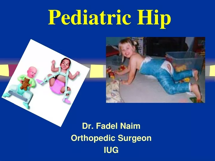
Pediatric Hip Dr. Fadel Naim Orthopedic Surgeon IUG
The main aim of this presentation is to: • emphasize the importance of the early identification of DDH • The earlier an abnormality of the infant hip is detected, the simpler and more effective the treatment will be
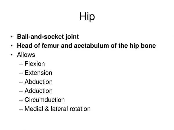
Developmental Dysplasia of the Hip (DDH) • DDH is a term used to describe a spectrum of abnormalities affecting the relationship of the femoral head to the acetabulum. • an immature hip • a hip with mild acetabular dysplasia • a hip that is dislocatable • a hip that is subluxated • a hip that is frankly dislocated.
Teratologic Hip : Fixed dislocation Occurrs prenatally Often with other anomalies • Dislocated Hip : Completely out May or may not be reducible • Subluxated Hip : Only partially in • Unstable Hip : Femoral head can be dislocated • Acetabular Dysplasia : Shallow Acetabulu Head Subluxated or in place
Epidemiology • 1 in 100 newborns examined have evidence of instability ( positive Barlow or Ortolani) • 1 in 1000 live births true dislocation • most detectable at birth in nursery • 60% stabilize in 1st week and 88% stabilize in first 2 months without treatment remaining 12% true dislocations and persist without treatment
Risk Factors associated with DDH • Breech Presentation • Family History of DDH (especially if in parent or sibling) • Female Baby (DDH is 4 X likely to occur in a female infant) • Postnatal Positioning
Risk Factors associated with DDH • Breech Presentation • Family History of DDH (especially if in parent or sibling) • Female Baby (DDH is 4 X likely to occur in a female infant) • Postnatal Positioning
Risk Factors associated with DDH • decreased intrauterine space • Large Baby (>4kg) • Overdue > 42 weeks • Oligohydramnios • Associated with Plagiocephaly, Torticollis and foot deformities • First born baby or multiple pregnancies
PHYSICAL EXAMINATION • The reliability of physical examination changes as the child grows, therefore examination techniques vary depending on the age of the child.
Diagnosis • Clinical risk factors • Physical exam • Ortolani Test • Barlow Test
Neonatal ExaminationOrtolani (reduction test) Feel a Clunk Not hear a click !
Ortolani and Barlow tests are accurate within the first 48 hours of birth and then become increasingly less accurate. • Thus Hip examination ideally should be performed within 48 hours • After 3 months of age, the Ortolani and Barlow tests may be unreliable • therefore additional means of examination, used in combination with the Ortolani and Barlow tests, are necessary
Older Infants (> 3 months of age) • restricted abduction at the hips • leg length discrepancy • asymmetrical thigh and gluteal skin folds
Restricted Abduction At The Hips • The most sensitive sign associated with DDH in the older infant. • The examination should be performed gradually and may need to be repeated a number of times • Normal range of motion at the hip is abduction to 60˚ or more, with range less than this suggestive of DDH.
Leg Length Discrepancy • Total leg length discrepancy should be assessed in prone with hips and knees extended • assessing for leg length discrepancy using the Galeazzi Test.
Asymmetrical Skin Folds • Asymmetrical skin folds alone do not constitute a diagnosis of DDH • However this information can be used in combination with other physical signs during assessment.
Bilateral Dislocation • Diagnosis more diffecult • Abduction may be decreased symmetrically with bilateral dislocations. • Galeazzi Test may be negative in bilateral dislocations • There may be no asymmetrical skin folds
Klisic test • An imaginary line between anterior superior iliac spine and great trochanter should point towards or above the umbilicus. • If dislocated will point below
Late Signs • In children who are walking • a limp may be present • the child may toe-walk on the affected side. • If DDH is present in both hips • increased lumbar lordosis • prominent buttocks • a waddling gait
Ultrasound • Morphologic and dynamic assessment • Indications controversial due to high levels of over-diagnosis • Currently not recommended as a routine screening tool other than in high risk patients
Ultrasound Femoral head Abductors Ilium
Ultrasound Femoral head Abductors Ilium
Ultrasound Femoral head Abductors Ilium
Ultrasound Graf’s alpha angle >60 = normal *line w/ ilium bisects head 50/50
If the child is 6 weeks to 5 months of age, Ultrasound (US) is generally the most appropriate imaging technique • If the child is 5 months or greater, X-ray is generally the most appropriate imaging technique • Between 4 and 6 months, US and X-ray are equally effective diagnostic tools
Radiographs Summary • Femoral head appears 4 - 7 months • Shenton’s line • Perkin’s and Hilgenreiner’s lines • Inferomedial quadrant • Acetabular index • Normal < 30 (Weintroub et al)
Radiological Diagnosis X-ray findings • Delayed appearance of ossific nucleus • Small ossific nucleus • Dysplastic acetabulum • Proximal displacement of femur • Increased acetabular index ( n=27, >30-35 dysplasia) • Disruption Shenton line
Radiography in out
Radiography 39o 27o
Imaging < 30 wnl
Treatment Options • Age of patient at presentation • Family factors • Reducibility of hip • Stability after reduction • Amount of acetabular dysplasia