Download
1 / 15
520 likes | 2.14k Views
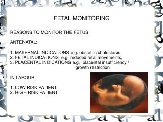
Electronic Fetal Monitoring. 2008 NICHD Workshop Definitions for Electronic Fetal Monitoring. Dione Ganser, BSN, RNC Walden University NURS 6320. objectives. Students will be able to -Discuss types of monitoring- external vs. internal. -Identify baseline FHR.

E N D
Electronic FetalMonitoring 2008 NICHD Workshop Definitions for Electronic Fetal Monitoring Dione Ganser, BSN, RNC Walden University NURS 6320
objectives Students will be able to -Discuss types of monitoring- external vs. internal. -Identify baseline FHR. -Identify FHR variability. -Identify FHR decelerations. -Identify FHR accelerations. -Understand reason for FHR declerations. -Discuss frequency, duration, and intensity of contractions.
History of electronic fetal monitoring Reference: Lyndon, A. & Ali, L.U. (2009), Sharma , D.L. (2010), Stevenson & Benitz (2003)
Monitoring contractions Palpation Internal Intrauterine Pressure Catheter External tocodynamometer
Monitoring fetal heart rate External devices for listening to fetus External continuous ultrasound monitors Fetoscope Early wooden stethescope Internal fetal scalp electrode for continuous fetal heart rate tracing. Doppler
Electronic Fetal monitors “Get me outta here!” –Love, Baby
Frequency of contractions are defined as the time in minutes between the start of one contraction and the start of the next. Contractions 1 minute 1 minute 1 minute 1 minute 2 minutes Intensity 60 seconds 1 minute 1 minute Intensity measures the strength of the contraction. IUPC is the only quantitative measurement of contractions. Duration is measured in seconds from the start to the end of a contraction.
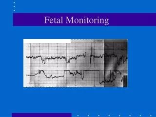
fhr baseline Baseline FHR is the median rate to the nearest 5 BPM. A normal FHR baseline is between 110 and 160 BPM. Below 110 BPM is bradycardia. Above 160 is tachycardia. 150 BPM 140 BPM Median 135 130 BPM 120 BPM The fetal heart rate in the above graph has visually apparent variability between approximately 130 and 140. The middle ground of this variability, to the nearest 5 BPM, is 135 BPM. Lyndon & Ali, 2009
Fhr variability Variability is the irregular amplitude fluctuations of the FHR. It is considered one of the most important predictors of adequate fetal oxygenation. Absent variability has undetectable amplitude. Minimal variability has FHR fluctuations between 1-5 BPM. Moderate variability has FHR fluctuations between 6-25 BPM. Marked variability has FHR fluctuations over 25 BPM. NCC Monograph, 2010
accelerations Fetal heart rate accelerations- Less than 32 weeks- 10 x 10 Greater than 32 weeks- 15 x 15 Acceleration Prolonged accelerations last for over 2 minutes but less than 10 minutes. An acceleration that last for more than 10 minutes is considered a baseline change. Prolonged accleration NCC Monograph
Variable deceleration This abrupt drop in FHR indicates umbilical cord compression. Variable decelerations are an abrupt drop in fetal heart rate regardless of relationship to contractions. NCC Monograph
Late deceleration The gradual change of late decelerations indicates utero-placental insufficiency. Late decelerations usually start after the peak of the contractions. This gradual decrease from baseline to nadir is over 30 seconds or more. These decelerations only occur with contractions. Intrauterine resuscitation measures- -Maternal repositioning -Fluid bolus -Correction of maternal hypotension -Evaluate and correct for tachysystole -Maternal oxygen supplementation by non-rebreather mask in the presence of minimal or absent variability. NCC Monograph
Early deceleration Early decelerations indicate fetal head compression. The gradual decrease of FHR mirrors the increase and decrease of contractions. Early Early NCC Monograph
Bradycardia Prolonged deceleration Tachycardia Pseudosinusoidal Sinusoidal Please find more helpful information at : dmganser2.weebly.com
references Fedorka, P. (2010). Electronic fetal monitoring: an update. Journal of Legal Nurse Consulting, 21(1), 15-18. Retrieved from EBSCO http://web.ebscohost.com.ezp.waldenulibrary.org/ehost/pdfviewer/pdfviewer?si d=e2f19c0f-c74c-41f2-b5cc-7b4eb9485962%40sessionmgr13&vid=7&hid=15 Lyndon, A. & Ali, L.U. (Eds.) (2009). Fetal Heart Monitoring: Principles and Practices (4th ed.). Washington D.C.: Kendall Hunt. Macones, G.A., Hankins, G.D.V., Spong, C.Y., Hauth, J., & Moore, T. (2008). The 2008 National Institute of Child Health and Human Development workshop report on electronic fetal monitoring. Journal of Obstetrics, Gynecologic and Neonatal Nursing, 37(5), 510-515. Murray, S.S. & McKinney, E.S. (2010). Foundations of maternal-newborn and women’s health nursing (5th Ed.). Maryland Heights, MO: Elsevier. National Certification Corporation (2010). NICHD Definitions and classifications: Application to electronic fetal monitoring interpretation. NCC Monograph, 3(1). Sharma, D.L. (2008). Electronic fetal monitoring [Slideshow]. Retrieved from http://www.obgyn.net/educational-tutorials/article/12571. Stevenson, D.K. & Benitz, W.E. (2003). Fetal and neonatal brain injury: Mechanisms, management, and the risks of practices (3rd Ed.). New York, NY: Cambridge University Press.