Download
1 / 36
400 likes | 972 Views
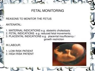
Fetal Monitoring. Introduction. 1600’s Kilian proposes the use of fetal heart rate to diagnose fetal distress 1893 criteria for determining fetal distress by Von Winckel Tachycardia >160bpm Bradycardia<100 Irregular heart rate Passage of meconium Alteration of fetal movement. Introduction.

E N D
Introduction • 1600’s Kilian proposes the use of fetal heart rate to diagnose fetal distress • 1893 criteria for determining fetal distress by Von Winckel • Tachycardia >160bpm • Bradycardia<100 • Irregular heart rate • Passage of meconium • Alteration of fetal movement
Introduction • EFM introduced in late 1950’s with first commercial product in 1968 as an alternative to auscultation • Initially utilized for high risk patients, but has become nearly universal • 44.6% of live births in 1980, increased to 62.2% in 1988 Albers and Krulewitch OB Gyn.1993;82:8-10. • Early observational studies suggested reduced perinatal mortality
Physiology • Fetal heart rate controlled by autonomic nervous system, with goal to maintain brain perfusion • Parasympathetic control increases with age, thus heart rate decreases with gestational age • Baroreceptors and chemoreceptors play a large role in the control of heart rate
Fetal Oxygenation • Placentation • Maternal hypotension • Microvascular disease (HTN, PIH, Diabetes, collagen vascular disease) • Cord factors--knot, nuchal cord, stretch, compression
DR C BRAVADO • Determine Risk • Contractions • Baseline RAte • Variability • Accelerations • Decelerations • Overall Assessment • ALSO Fourth Edition
Baseline Rate • Normal between 120-160 (110-160) under vagal control (if give atropine increase HR to 160) • Tachycardia • Mild 160-180 • Severe>180 • Bradycardia • mild 100-120 • severe <80
Causes of Tachycardia • Hypoxia • Infection • Maternal hyperthyroidism • Fetal anemia • Fetal Heart Failure • Fetal cardiac tachydysrhythmia • Drugs (vagolytic and sympathomimetic)
Causes of Bradycardia • Hypoxia/acidosis • Hypothermia • Fetal cardiac bradydysrhythmia • Heart block (SLE) • Drugs • False bradycardia (maternal tracing)
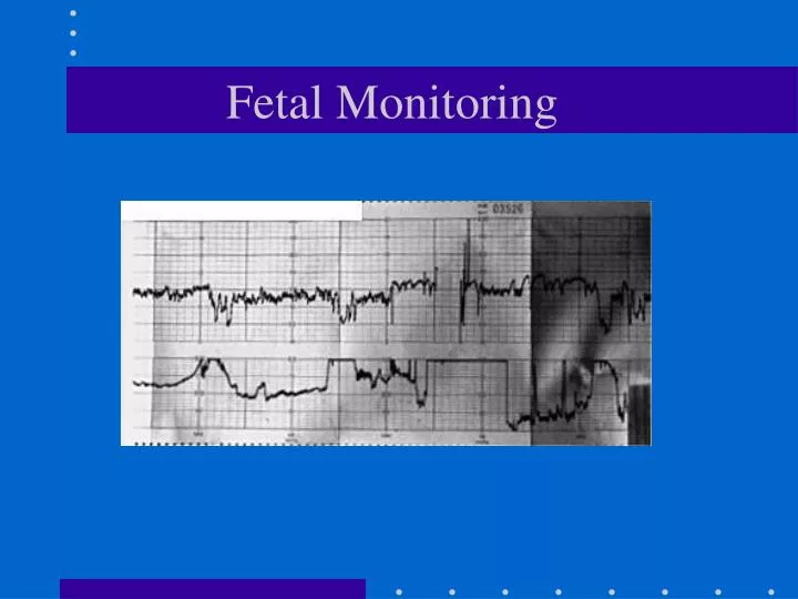
Variability • Short term--instantaneous changes from beat to beat • Long term beat to beat--variability over the course of a minute (the waviness of the pattern) • 1997 NICHD (National Institute of Child Health and Human Development) Fetal Monitoring Workshop did not recommend differentiating short and long term variability
Variability Classification • Absent • minimal < 5 bpm variability • normal 6-25 bpm variability • marked >25 bpm variability
Acidosis/hypoxia Congenital abnormalities (CNS) Sleep cycles Prematurity Tachycardia Sepsis Damaged CNS Drugs Narcotics Demerol--decreased BTBV in 5 min and lasts for about 1 hr or longer Barbiturates General anesthesia Parasympatholytics Phenothiazine Causes of decreased BTBV
Acceleration • Change in heart rate above the baseline • Usually use 15 bpm above baseline for 15 sec. (initially developed for non stress testing)
Decelerations • Early deceleration • Variable deceleration • Late deceleration • Prolonged deceleration
Early Deceleration • Head compression with altered cerebral blood flow causes vagal stimulus • U shaped with nadir coinciding with peak of contraction • Return to baseline by the end of the contraction • Rarely < 100-110bpm or 30-40bpm below baseline • Occur at 4-7 cm dilation
Variable Decelerations • Variables occur in 50-80% of labors during 2nd stage • Variable timing, shape, depth • Onset is abrupt as is the return to baseline • Caused by cord compression, or spasm as cord stretched • Occlusion of the vein reduces blood return, hypotension stimulates the baroreceptors increasing the heart rate • Occlusion of the artery increases vascular resistance and blood pressure causing a baroreceptor mediated deceleration in heart rate • Concerning if late in timing, duration >2 minutes, slow return with late component, lose shoulders,
Variable Decelerations • Mild Variable-greater than 80 bpm, or last less than 30 sec. in duration regardless of depth • Moderate Variable-deceleration to < 80 bpm • Severe Variable- deceleration to <70 for >60 secs
Late Decelerations • Always represent hypoxia • Oxygen sensors increase vascular tone, leading to baroreceptor mediated deceleration • Myocardial depression also plays a role • Smooth symmetric decrease in heart rate at or after peak of contraction return to baseline after end of contraction • Rarely more than 30-40 bpm drop (usually 10-20)
Late Decelerations • Animal studies--the shorter the onset of late after contraction the worse the O2 sat • Difficult to determine level of acidosis by depth of deceleration • Duration of repetitive late deceleration impacts acidosis
Maternal hypotension Hyperactivity of the uterus often iatrogenic Chronic hypertension Preeclampsia Collagen Vascular diseases Maternal diabetes Maternal hypoxia resulting in hypoxemia Maternal severe anemia Fetal anemia Late Deceleration
Prolonged Deceleration • Isolated deceleration lasting 90-120 seconds or more (2-10 minutes by others) • Multiple mechanisms, but profound stimuli • Concerning if slow return to baseline, rebound tachycardia, loss of variability
Prolapsed cord Post epidural hypotension Prolonged cord compression Uterine tetany Severe abruption Eclampsia Rapid descent in the birth canal Paracervical block Prolonged scalp stimulation as in placement of FSE Prolonged Deceleration
Other Patterns • Hypervariability or saltatory--Sign of hypoxia • Sinusoidal pattern--regular sine wave pattern about 2-5 cycles per minute lasting at least 2 minutes with amplitude 5-15bpm with loss of BTBV • Sign of severe fetal anemia and/or hypoxia • Pseudosinudoidal--varies in shape and amplitude and BTBV maintained
Benefits May decrease infantile seizure rate Am J OB Gyn 1985;152:524-539. Does not require nurse to be at the bedside Risks Risks and Benefits
Benefits May decrease infantile seizure rate Am J OB Gyn 1985;152:524-539 Risks Does not require nurse to be at the bedside Risks and Benefits
Benefits May decrease infantile seizure rate Am J OB Gyn 1985;152:524-539 Risks Does not require nurse to be at the bedside Limits mobility Shown to increase instrumentation and cesarean rates without improvement in morbidity and mortality Trauma from internal monitors Risks and Benefits
Cardiotocography versus AuscultationBMJ 2001;322:1457-1462 • Inclusion criteria: Presented to the hospital and were followed in a hospital or community based clinic • Exclusion criteria: PIH, HTN, DM, IUGR, previa, abruption, vaginal bleeding, fetal anomaly, VBAC, Rh disease, breech, multiple gestation • Randomized at an outpatient appointment to 20 minutes Cardiotocography vs. doppler for at lease one contraction
Cardiotocography versus AuscultationBMJ 2001;322:1457-1462 • Outcomes • Primary: Metabolic acidosis • Secondary: Apgar, ventilation, NICU admission, obstetric intervention
Cardiotocography versus AuscultationBMJ 2001;322:1457-1462 • Results • 3752 women randomized • Umbilical artery pH <7.2 OR 0.96 (0.79-1.17) • Apgar at 5 minutes <7 OR 1.07 (0.65-1.75) • Use of scalp pH OR 1.14 (0.91-1.42) • CLE use OR 1.15 (1.00-1.32) • Caesarian OR 1.20 (0.96-1.50) • Operative delivery OR 1.15 (1.00-1.32)
Cardiotocography versus AuscultationBMJ 2001;322:1457-1462 • Subgroup analysis with 1736 who remained low risk • Umbilical artery pH <7.2 OR 1.02 (0.79-1.31) • Apgar at 5 minutes <7 OR 1.39 (0.72-2.66) • CLE use OR 1.33 (1.10-1.61) • Caesarian OR 1.43 (0.95-2.18) • Operative delivery OR 1.36 (1.12-1.65)
ACOG Guidelines for High Risk Patients • During the active phase of the first stage of labor, when intermittent auscultation is used, the FHR should be evaluated and recorded at least every 15 minutes following a uterine contraction. If continuous EFM is used, the tracing should be evaluated at least every 15 minutes • During the second stage of labor, the FHR should be evaluated and recorded at least every 5 minutes when auscultation is used and should be evaluated at least every 5 minutes when EFM is used