Download
1 / 28
410 likes | 1.21k Views
Fetal Monitoring. Ultrasonography. Uses reflection of very high frequency sound waves of between 3.5 to 7.0 megahertz (i.e. 3.5 to 7 million cycles per second). Monitoring: Chorionic sac during embryonic period placental and fetal size multiple births abnormal presentations

E N D
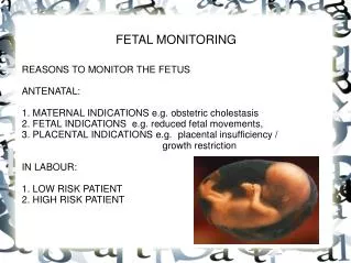
Ultrasonography Uses reflection of very high frequency sound waves of between 3.5 to 7.0 megahertz (i.e. 3.5 to 7 million cycles per second) Monitoring: • Chorionic sac during embryonic period • placental and fetal size • multiple births • abnormal presentations • biparietal diameter
Fetal Blood Sampling Usually from the scalp, fetal blood pH is a good indicator of placental gas exchange. In the past, fetal blood sampling was used only during labor through the mother's open cervix to test blood from the fetal scalp for oxygenation. Today, in many perinatal care centers, fetal blood sampling is performed by specially trained perinatologists as part of diagnosing, treating, and monitoring fetal problems at various times during pregnancy.
Fetal Blood Sampling A fetal blood sample may be taken to: • diagnose genetic or chromosome abnormalities. • check for and treat severe fetal anemia or other blood problems such as Rh disease. • check for fetal oxygen levels. • check for fetal infection. • give certain medications to the fetus.
How is fetal blood sampling performed? A long, thin needle is inserted into the mother's uterus guided by ultrasound. Blood may be taken from several sources: • blood vessels of the umbilical cord (also called cordocentesis, funicentesis, or percutaneous umbilical blood sampling, or PUBS) • a fetal blood vessel, usually in the liver or heart Fetal blood transfusions may also be performed in this
Amniocentesis also referred to as amniotic fluid test or AFT • used in prenatal diagnosis of chromosomal abnormalities and fetal infections, in which a small amount of amniotic fluid, which contains fetal tissues, is extracted from the amnion or amniotic sac surrounding a developing fetus, and the fetal DNA is examined for genetic abnormalities. • Little amniotic fluid present prior to 12th week of gestation
Chorionic Villus Sampling • chromosomal abnormalities etc. • The advantage of CVS is that it can be carried out 10-13 weeks after the last period, earlier than amniocentesis (which is carried out at 16-20 weeks).
Alpha-Fetoprotein Assay • AFP is a glycoprotein synthesized in the fetal liver and yolk sac. • The fetus normally excretes AFP into its urine, hence into the amniotic fluid. • High levels may also be present due to: • open neural tube defect • open abdominal wall defect • skin disease or other failure of the interior or exterior body surface. • Various forms of tumours
Placental Insufficiency • Placental defects effectively reduce available surface area • reduced uteroplacental blood flow may also occur due to maternal hypotension or renal disease.
Multiple Pregnancy • Individuals of multiple births usually weigh considerably less • in the third trimester placenta may not be able to supply the total requirements for multiple births
Small Babies • Low birth weight: • < 2,500g • Premature: • < 37 weeks of gestation • Small for Date: • Smaller than expected for age
Gradient vs Indicator • Maturity Gradients • assessment of the relative rates of development of parts or structures of the body • Maturity Indicators • Some characteristic of the body that has distinct stages of development that all normally developing children will pass through
Bigness vs Maturity • Do not confuse size with maturation • Obesity often associated with advanced maturation skeletally but not in muscular development
Maturity Indicators • Age of Peak Height Velocity • Skeletal Age • Dental Age • Menarche • Secondary Sexual Characteristics
Closer relationship of Age at Menarche with Skeletal Age (SA) than Chronological Age (CA)