Download
1 / 29
290 likes | 348 Views
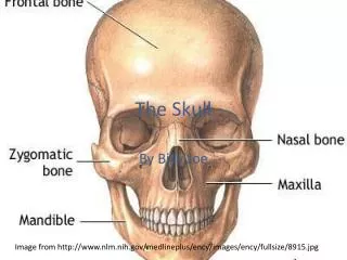
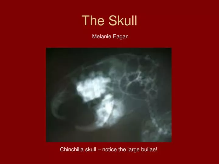
The Skull. Melanie Eagan. Chinchilla skull – notice the large bullae!. Indications for skull rads. Neurological problems Nasal problems Mandibular problems Maxillary problems 1 ̊ tumors of skull Mass behind eye Teeth diseases Middle ear problems. Views.

E N D
The Skull Melanie Eagan Chinchilla skull – notice the large bullae!
Indications for skull rads • Neurological problems • Nasal problems • Mandibular problems • Maxillary problems • 1̊ tumors of skull • Mass behind eye • Teeth diseases • Middle ear problems
Views • Careful positioning is necessary • Sedation or GA usually necessary • Positioning aids to elevate cassette
Intraoral dorsoventral view • Good for rostral aspect of nasal cavities
Ventrorostral-dorsocaudal oblique • Good for more caudal aspect of nasal cavities • More difficult to assess rostral aspect of nose (shortened by angulation of x-ray beam)
Rostrocaudal view • Patient positioning for rostrocaudal rads of frontal sinuses taken with vertical beam
Rostrocaudal view • Used for viewing frontal sinuses • Rotation of head must be avoided so view is not “obliqued” • Open mouth rostrocaudal used to view tympanic bullae and foramen magnum
Lateral oblique used to view: • Tempromandibular joint • Teeth in mandible/maxilla • Fractures in mandible/maxilla
differences in the cat skull Dog skull – left Cat skull – right Arrow pointing to cribiform plate • Cats have: • greater doming on frontal and nasal bones • smaller frontal sinuses (may be absent in Persians) • more complete bony orbits • wider skulls ( due to wider zygomatic arches)
Rads or CT? • CT • Elimination of superimposition • Ability to display images in multiple planes • Shorter imaging time • Higher contrast resolution • Higher cost • Lower availability • CT and rads both underestimate presence of mild middle ear disease • CT more consistent for moderate/severe middle ear disease
nasopharyngeal polyps in cats • Polyps: • Benign growths • Nasopharynx, middle ear, external ear canal • Diagnostic Imaging: • Rads of skull with emphasis on tympanic cavities • Lateral oblique and open mouth views to see changes in tympanic bullae (normally contain air) • Rad changes suggesting polyps: • Soft tissue densities in bullae • Evidence of chronic otitis media (bony thickening)
Nasopharyngeal polyps • Rads: only partially sensitive diagnostic tool for otitis media • 25% of animals with middle ear disease have no radiographic abnormalities • CT or MRI: • Define extent of mass in middle ear • Determines invasion into inner ear, pharynx, outer ear more clearly than rads
Other Skull Problems • Tempromandibular joint disease • Canine craniomandibular osteopathy • TMJ dysplasia • Luxation/subluxation • Fracture • Ankylosis • Otitis • Externa • Media • Interna • Tumors
TMJ TMJ “hinge joint” – condyloid process of mandible articulates with mandibular fossa of temporal bone
DV of left TMJ M= mandible PC=coronoid process of ramus of mandible Z= zygomatic arch C= condyloid process of mandible a = angular process of mandible F= mandibular fossa P= articular process of temporal bone Between arrowheads = thin, radiolucent TMJ space
Canine CranioMandibular Osteopathy • Unknown etiology • Common in West Highland, Scottish, and Cairn terriers • Extensive, bilateral, irregular, periosteal reaction of mandible • Extend to TMJ, tympanic bullae, calvarium • Rads to investigate TMJ • Dogs have difficulties opening mouth during mastication
TMJ Luxation • Consequence of trauma, dysplasia, degeneration, idiopathic condition • Condylar process of mandible luxated rostrodorsally • Dental malocclusion present • Unilateral luxation w/ mandibular fx (dogs) • Unilateral luxation with or without mandibular fx (cats)
TMJ luxation Mandibular fossa of temporal bone is not articulated with condyloid process of mandible. The condyloid process has rotated forward and upward.
TMJ ankylosis • Relatively uncommon or undiagnosed • Abnormal immobility and consolidation of a joint • Consequence of untreated intra-articular (trueankylosis) or extra-articular (false ankylosis) trauma • Hemarthrosis syspected as initiating factor • Cat falling from great height • From extensive new bone formation • otitis media or canine craniomandibular osteopathy
TMJ ankylosis Transverse CT image: Bilateral true ankylosis
TMJ tumors • Most common: • Osteosarcoma • Multilobular osteochondrosarcoma • Characteristic appearance on rads, CT, MRI • Rounded, well defined, osseous mass • Course, granular architecture arising from mandible, zygomatic arch or other flat bones of skull
TMJ tumor Transverse (A) and Dorsal (B) plane images: lobulated bony mass arising from left maxilla and zygomatic bone with compression (not invasion) of adjacent bone. Characteristic of multilobular osteochondrosarcoma.