Download
1 / 32
330 likes | 651 Views
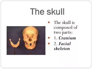
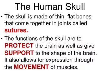
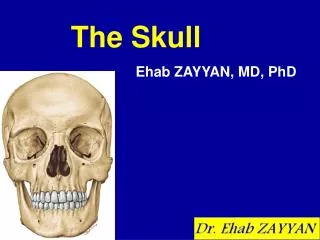
The Skull. There are 22 bones in the human skull. Divided into two groups, the cranial vault and the face. The cranial vault contains 8 bones. These directly surround and protect the brain. The face contains 14 bones. 13 of which form the major structure of the face. The mandible is 1 bone.

E N D
There are 22 bones in the human skull. • Divided into two groups, the cranial vault and the face. • The cranial vault contains 8 bones. These directly surround and protect the brain. • The face contains 14 bones. 13 of which form the major structure of the face. • The mandible is 1 bone. • There are three bones found in each inner ear.
Cranial bones Frontal (1) Sphenoid (1) Parietal (2) Ethmoid (1) Occipital (1) Temporal (2)
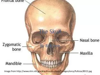
Frontal bone • Forms anterior portion of skull. • Upper margin contains supraorbitalforamen. • Also contains two frontal sinuses.
Parietal • One on each side of skull behind frontal bone. • Fused together at the midline along sagittalsuture. • Join frontal bone at the coronalsuture.
Occipital • Joins the parietal bones along the lambdoidal suture. • Forms the back of the skull and base of the cranium. • Foramenmagnum found on lower surface. • Occipitalcondyles (on either side of foramen magnum articulate with C – 1.
Temporal • One on each side of skull. • Joins parietal bone along the squamosalsuture. • Externalacousticmeatus located near inferior margin. • Mandibularfossae articulate with mandibularcondyles.
Temporal bone continued • Mastoidprocess and styloidprocess both project from the temporal bone. • The zygomaticprocess projects anteriorly from the temporal bone, and joins the zygomaticbone.
Sphenoid bone • Found in the anterior cranium. • Central part and two “wings” extending laterally toward each side of the skull. • Forms base of cranium, sides of the skull and the floor and sides of the orbits. • Central portion contains sellaturcica, where the pituitary gland sits. • Contains sphenoidalsinus.
Ethmoid bone • In front of sphenoid. • Two sides joined by cribriformplate. • Plate forms roof of nasal cavity. • Perpendicular plate projects down from middle of cribriform plate and forms nasal septum. • Contains curled plates called conchae, superior and middle. • Contains ethmoidalsinuses.
Maxillae • Forms upper jaw. • Compose anterior roof of mouth, floors of orbits and sides and floor of nasal cavity. • Also contain sockets of upper teeth. • Maxillary sinuses. • Palatine processes grow together to form anterior portion of hard palate.
Palatine bones • L-shaped • Located behind maxillae • Horizontal part forms posterior of hard palate and floor of nasal cavity. • Perpendicular portion forms lateral walls of nasal cavity.
Zygomatic bones • Form the prominences of the cheek below and to the sides of the eyes. • Form lateral walls and floors of the orbits. • Has a temporal process extending posteriorly to join zygomatic process of temporal bone. • This form zygomaticarch.
Lacrimal bones • Thin, scalelike structure located on the medial orbit wall between ethmoid and maxilla.
Nasal bones • Long and thin. • Lie side by side and are fused at the midline forming the bridge of the nose.
Vomer bone • Thin and flat. • Midline of nasal cavity • Joins perpendicular plate (posteriorly) and forms nasal septum.
Inferior nasal conchae • Scroll shaped bones. • Attached to lateral walls of nasal cavity • Support mucous membranes within the nasal cavity.
Mandible • Horizontal horse-shoe shaped bone with processes projecting upward at each end. • This projection is divided into the posterior mandibular condyle and anterior coronoid process. • Coronoid process provides attachments for muscles used in chewing. • Alveolar arch contains hollow sockets for lower teeth.