Download
1 / 66
1.36k likes | 3.07k Views
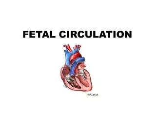
Fetal biometry. The common tomograms used : Axial Coronal Sagittal (midline). F etal biometry . Fetal growth can be monitored accurately later in life only if the exact information about the GA is available. As less than 50% of women are certain about their LMP.

E N D
The common tomograms used : • Axial • Coronal • Sagittal (midline)
Fetal biometry • Fetal growth can be monitored accurately later in life only if the exact information about the GA is available. • As less than 50% of women are certain about their LMP. • Menstrual cycle is not 28days long • Irregular • Taking COC • Women had bleed in early pregnancy • Lactating women
Gestational age: length of the pregnancy based upon reliable LMP, assuming that conception occurs 14 days later. • Postmenstrual age: the length of pregnancy based on the LMP, irrespective of its reliability.
Several US parameters have been used to estimate GA, the most commonly used are: Mean sac diameter Gestational sac volume Crown rump length Biparietal diameter Femur length
Crown rump length • The biologic variability of CRL is small & growth is very rapid. • However there are still a number of factors that can affect the size of embryo; • Measurement errors • Different in growth rate between individuals • Fetal sex • Maternal conditions • CRL may indicates an early IUGR.
Tend to underestimate GA by 2-3days After 12wa CRL m inaccuracy of 7-10days
Crown – rump length • To establish correct GA : Unflexed Longitudinal section The end point of the crown & rump clearly defined Placing the calipers correctly on this defined end point.
Crown rump length • CRL between 5-7ws are incorrect: • The very early embryo is unflexed. • The full length of the embryo has not been obtained. • The end point of the embryo is closely adjacent to yolk sac or wall of GS.
Crown rump length • After 7ws its easily to identify the end points of the embryo, but insure that you are imaging the maximum length of the embryo. • The CRL should be measured from 3 different images and the measurements should be agree to within 3mm in the embryo & 5mm in the fetus.
CRL measurement problems • Any degree of flexion of fetal spine will produce an underestimate of CRL when linear calipers are used.
CRL measurement problems • When the fetus remains obstinately curled, you have 4 choices: • Sit and wait. • Measure the flexed length using onscreen nonlinear measuring facilities. • Use the linear caliper to measure the parts of the fetal length that are in straight sections and add them together.
4-Using a linear calipers along the flexed length. This is not to be recommended under any circumstances How easy to produce errors of 10-15mm simply by measuring 12-13ws fetus incorrectly
2nd trimester biometry- –assessing gestational age
2nd trimester biometry –assessing gestational age • BPD & FL provide the most accurate assessment of GA. • HC, TCD & AC they provide further confirmation of GA and aid in the exclusion of growth related abnormalities.
BPD • The BPD has traditionally been the most widely used ultrasound parameter in the estimation of gestational age • - A single optimal measurement of the BPD will predict the gestational age to within ± 5 days.
Biparietal diameter (BPD) • BPD : maximum diameter of transverse section of the fetal skull at the level of parietal eminence . • BPD, OFD & HC can be measured from: Lateral ventricles view Thalami view
Lateral ventricles view of BPD • A rugby- football- shaped skull, rounded at the back (occiput) and more pointed at the front (synciput). • Long midline equidistant from the proximal and distal skull.
Lateral ventricles view of BPD • The CSP bisecting the midline 1/3 of the distance from the synciput to the occiput. • The two ant horn of lateral ventricles placed about the midline. • The two post horn of lateral ventricles placed about the midline
Trans thalamic view of BPD A rugby- football- shaped skull, rounded at the back (occiput) and more pointed at the front (synciput). Short midline equidistant from the proximal and distal skull
Trans thalamic view of BPD • The CSP bisecting the midline 1/3 of the distance from the synciput to the occiput. • The thalami • The basal cisterns.
a b
Measurement • Outer to outer!!!!! • Outer to inner!!!!!!!
Trans cerebellar diameter It’s a beast dater of pregnancy. TCD in mm= ws of gestation until 22ws.
Suboccipitobregmatic view • M. at 90 degree with the longest axis • The bonus with TCD is that it force the operator to image the entire post fossa which indirectly refractor of the integrity of neural tube. • The same plane of nuchal fold thickness.
Measurement of HC • Short midline, 90 degrees to the beam • Oval shape • Thalami • NO cerebellum or orbits • Cavum septum pellucidi • Measure circumference of outer bone (usual to put calipers at occiput then sinciput)
Abdominal circumference • The AC is measured in a location that estimates liver size. • The liver is the largest organ in the fetal torso, and its size reflects aberrations of growth, both growth restriction and macrosomia.
A circular section of the abdomen ,unbroken & short rib echo of = size on each side. • A cross-section of one vertebra visualized as a triangle of 3 white spots. • A short length of umbilical vein (1/3 from ant abd wall to spine). • The stomach in the left side of the abd. • NO kidney, bladder, heart. Adrenal allowed
A. This plane is too caudal B. This is the correct level for AC. C. this plane is too inclined in a craniocaudal axis.
Femur length • 12ws to term, The upper femur , Full length of femur • Soft tissue should be visible beyond both ends of the femur. • The bone should not appear to emerge with the skin of the thigh at any point.
Femur length • The FL measurement is made from the center of the U shape at each end of the bone. • Good practice, to obtain measurements from separate image of the same femur. (1mmof each other).
Cephalic index= BPD/OFD=80± 5 Dolichocephaly ( narrow BPD) ,75 Brachycephaly (wide BPD)
2nd trimester fetal anatomy • Intracranial anatomy. • Nuchal area. • Fetal spine • Fetal chest & diaphragm. • Fetal heart • Fetal abdomen • Urinary tract. • Fetal limbs • Fetal face. • Fetal sex
Intracranial anatomy • Cerebral ventricles • Thalami & 3rd ventricle • Cerebellum • Cisterna magna
Choroid plexus cyst • <10mm in diameter. • Unilateral or bilateral. • Resolve by 26ws • 0.5-2%
Cerebellum Cisterna magna