Download
1 / 32
330 likes | 473 Views
Chapter 44 Neurons and Nervous Systems. Biology 102 Tri-County Technical College Pendleton, SC. Functions of Nervous System. Sensory input is conduction of signals from sensory receptors to integration centers

E N D
Chapter 44 Neurons and Nervous Systems Biology 102 Tri-County Technical College Pendleton, SC
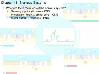
Functions of Nervous System • Sensory input is conduction of signals from sensory receptors to integration centers • Integration is process by which info from environmental stimulation of sensory receptors is interpreted and associated with appropriate responses of the body • Carried out mostly in central nervous system (CNS) which in vertebrates includes the brain and spinal cord
Functions, cont. • Motor output is conduction of signals from integration center (CNS) to effector cells (muscle cells or glands that actually carry out body’s responses to stimuli • **Nerves that communicate motor/sensory signals between CNS and rest of body are collectively called the peripheral nervous system (PNS) • Nerve defined as ropelike bundles of extensions of neurons tightly wrapped in connective tissue
Only have one neuron left and… • Functional unit of nervous system is the neuron (cell specialized for transmitting chemical and electrical signals from one location in body to another • Large cell body contains most of cytoplasm, nucleus, and other organelles • Cell bodies of most neurons located in CNS • Some neurons have cell body located in ganglia outside the CNS
Neuron, cont. • Dendrites: Fiberlike extension (process) that conveys impulse TO the cell body • Short, numerous, and extensively branched to increase surface area where cell most likely to be stimulated • Axons: the other type of extension (process) that is specialized to conduct signals AWAY from the cell body
Neuron III • Axon is long, simple process • Vertebrate axons in PNS wrapped in concentric layers of Schwann cells which form insulating myelin sheath • In CNS, myelin sheath formed by oligodendrocytes • Axons extend from axon hillock (where signals generated) to many branches tipped with synaptic terminals that release neurotransmitters
Neuron IV • Synapse is gap between synaptic terminal and target cell/dendrites of another neuron/or an effector cell • Neurotransmitters are chemicals that cross the synapse to relay the impulse
Glial Cells • Supporting cells (glia cells) structurally reinforce, protect, insulate, and assist neurons • Do NOT conduct impulses • Outnumber neurons 10x-50x • Several classes of glia are present • Astrocytes encircle capillaries in brain and contribute to blood-brain barrier • Restricts passage of most substances into CNS • Communicate with each other/other neurons via chemical signals
Glial Cells, cont. • Oligodendrocytes form myelin sheaths that insulate CNS nerve processes • Schwann cells form insulating myelin sheath around axons in PNS • Myelination provides insulating myelin sheath & increases speed of nerve impulse propagation • Microglia are phagocytes that dispose of debris (dead brain cells, bacteria, and the like) • Ependymal cells line cavities of brain and spinal cord where their beating cilia help circulate CSF
Rest? What’s rest? • All living cells have electrical charge difference across their plasma membrane • This difference gives rise to electrical voltage gradient across the membrane • Voltage measured across plasma membrane is called membrane potential • Typically ranges from minus 50 to minus 100 mV in animal cells
Resting Potential, cont. • Voltage outside cell called “zero” • Minus sign indicates inside of cell is negative in charge with respect to outside • For “resting” neuron (not transmitting an impulse), membrane potential of –70 mV is typical • Membrane potential of excitable cell at rest (unexcited state) is called resting potential
Action Potential • AP is rapid change in membrane potential of excitable cell that is caused by stimulus-triggered selective opening and closing of voltage-gated ion channels • AP is all-or-none event • Selective permeability of PM maintains ionic differences • Ions (charged molecules) cannot readily diffuse through hydrophobic core of PM’s phospholipid bilayer
Action Potential, cont. • Ions can cross membranes by carrier-mediated transport of by passing through ion channels • Ion channel is integral transmembrane protein that allows specific ion to cross membrane • May be passive (open all time) or gated (requires stimulus)
Action Potential III • Presence of gated channels in neurons permits excitable cells to change PM’s permeability and alter membrane potential in response to stimuli received by cell • K+ and Na+ • Depolarizing threshold potential usually around –50 to –55 mV
AP Propagation • AP is localized electrical event…a membrane depolarization at a specific point of stimulation • Neuron usually stimulated at dendrites or cell body • For AP to function as signal, it must somehow “travel” along axon to other end of cell • AP is regenerated ANEW in sequence along the axon
AP Propagation, cont. • AP is like tipping over row of standing dominoes • Strong depolarization of one AP assures neighboring region of neuron will be depolarized above threshold, triggering new AP at that location and so on to end of the axon…
Synaptic Transmission • Synapse is tiny gap between synaptic terminal of axon and signal-receiving portion of another neuron/effector cell • Presynaptic cell is transmitting cell • Postsynaptic cell is receiving cell • Two types of synapses: ELECTRICAL and CHEMICAL
Synapses, cont. • Electrical synapse allows action potentials to spread directly from pre-to postsynatpic cells via gap junctions (intercellular channels) • Allows impulses to travel from one cell to next without delay or loss of signal strength • Electrical synapses are MUST less common than chemical synapses
Synapses III • At chemical synapse, synaptic cleft separates pre-and postsynaptic cells • NOT electrically coupled • Synaptic vesiclesrelease neurotransmitter moleculesreleased into synaptic cleft by exocytosisdiffuse to postsynaptic membrane where binds to specific receptorscauses ion gates to open
Synapses IV • Neurotransmitter may excite or inhibit postsynaptic cell • Excite causes depolarization; inhibit causes hyperpolarization • Neurotransmitter quickly degraded by enzymes • Allows transmission in ONLY 1 direction
Neurotransmitters • Acetylcholine is major neurotransmitter • Degraded by acetylcholinesterase • Anyone care to spray a bug (organophosphates) • enough said • Other neurotransmitters are biogenic amines (epinephrine, norepinephrine, dopamine, histamine, and serotonin); amino acids (glutamate, glycine, asparatate, and gamma-aminobutyric acid [GABA]); neuropeptides (endorphins, enkephalins, and Substance P); gas (nitric oxide and carbon monoxide); purines (ATP and adenosine)
EPSP and IPSP • Excitatory postsynaptic potentials (EPSP) occur when excitatory synapses release a neurotransmitter that opens gated channels allowing Na+ to enter cell and K+ to leave (depolarization) • Inhibitory postsynaptic potentials (IPSP) occur when neurotransmitters released from inhibitory synapses bind to receptors that open ion gates which makes membrane MORE permeable to K+ (which leaves the cell) and/or to Cl- (which enters cell) casuing hyperpolarization
EPSP and IPSP, cont. • EPSPs and IPSP are graded potentials • They vary in magnitude with number of neurotransmitter molecules binding to postsynaptic receptors
To fire or not to fire…? • Single EPSP rarely strong enough to trigger an action potential • Additive effect (summation) from several terminals or repeated firing of terminals can change membrane potential • Temporal summation: chemical transmissions from 1 or more synaptic terminals occurs so close in time that each affects membrane while it is partially depolarized and before it has returned to resting potential
Summation, cont. • Spatial summation: several different synaptic terminals, usually from different presynaptic neurons, stimulate postsynaptic cell at same time and have additive effect of membrane potential • EPSPs and IPSPs can summate, each countering the effects of the other
Summation III • At any instant, axon hillock’s membrane potential is average of summated depolarizations due to all EPSPs and the summated hyperpolarizations due to all IPSPs • AP is generated when EPSP summation exceeds IPSP summation to point where membrane potential of axon hillock reaches threshold voltage
Neuropeptides • Neuropeptides often operate via signal-transduction pathway • Substance P is key excitatory signal that mediates perception of pain (humans) • Endorphins function as natural analgesics, decreasing perception of pain by CNS • Also decrease urine output, depress respiration, produce euphoria, and have other emotional effects • Endorphin also released from anterior pituitary as hormone affecting specific regions of brain (overlap between endocrine and nervous system)