Download
1 / 26
270 likes | 298 Views
Neurons and the Nervous System. Nervous System. Central nervous system (CNS): Brain Spinal cord Peripheral nervous system (PNS): Sensory neurons Motor neurons (somatic and autonomic). The Nervous System. The Nervous System. Central Nervous System (CNS). Peripheral Nervous System (PNS).

E N D
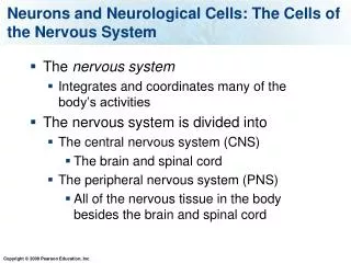
Nervous System • Central nervous system (CNS): • Brain • Spinal cord • Peripheral nervous system (PNS): • Sensory neurons • Motor neurons (somatic and autonomic)
The Nervous System The Nervous System Central Nervous System (CNS) Peripheral Nervous System (PNS) Brain Spinal Cord Motor Neurons Sensory Neurons • Somatic Nervous System • voluntary movements via skeletal muscles • Autonomic Nervous System • organs, smooth muscles Sympathetic - “Fight-or-Flight” responses Parasympathetic - maintenance
Divisions of the autonomic nervous system Rest Action
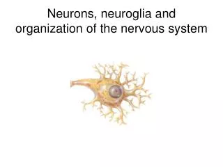
The Nervous System • A physical organ system like any other • The main cell of the nervous system are: • Neurons
The Neuron • The basic functional unit of the nervous system. • Function: Send impulses to and from the CNS and PNS
Dendrite Fine hair-like extensions on the end of a neuron. • Function: receive incoming stimuli. • Cell Body or Soma The control center of the neuron. • Function: Directs impulses from the dendrites to the axon. • Nucleus Control center of the Soma. • Function: Tells the soma what to do.
Axon Pathway for the nerve impulse (electrical message) from the soma to the opposite end of the neuron. • Myelin Sheath An insulating layer around an axon. Made up of Schwann cells. • Nodes of Ranvier Gaps between schwann cells. • Conduction of the impulse. (Situation where speed of an impulse is greatly increased by the message ‘jumping’ the gaps in an axon).
Types of Neurons • There are 3 types of neurons. • Sensory Neurons Neurons located near receptor organs (skin, eyes, ears). • Function: receive incoming stimuli from the environment. • Motor Neurons Neurons located near effectors (muscles and glands) • Function: Carry impules to effectors to initiate a response. • Interneurons Neurons that relay messages between other neurons such as sensory and motor neurons. (found most often in Brain and Spinal chord).
Nerves • Nerves Collections of neurons that are joined together by connective tissue. • Responsible for transferring impulses from receptors to CNS and back to effectors.
Three main types of neurons • Sensory Neurons • Interneurons • Motor Neurons
Sensory vs. Motor sensory nerve CNS e.g., skin Neurons that send signals from the senses, skin, muscles, and internal organs to the CNS motor nerve CNS e.g., muscle Neurons that transmit commands from the CNS to the muscles, glands, and organs Gray’s Anatomy 38 1999
Cell Body Dendrites MyelinSheath Axon Neurons Dendrites of another neuron Axon of anotherneuron
Neural Anatomy • Dendrite • the bushy, branching extensions of a neuron that receive messages and conduct impulses toward the cell body • Axon • the extension of a neuron, ending in branching terminal fibers, through which messages are sent to other neurons or to muscles or glands
Neural Anatomy and communication • Synapse • junction between the axon tip of the sending neuron and the dendrite or cell body of the receiving neuron • tiny gap at this junction is called the synaptic gap or cleft • Synapse movie
Specific Parts: The NeuronFunction 1. 3. 2. Neurons = 3 functions: Reception, Conduction, Transmission
Communication • Impulse releases neurotransmitter from vesicles • Neurotransmitter enters synaptic gap • Neurotransmitter binds to receptors on the receiving neuron
Myelin Sheath • Fatty material made by glial cells • Insulates the axon • Allows for rapid movement of electrical impulses along axon • Nodes of Ranvier: gaps in myelin sheath where action potentials are transmitted • Multiple sclerosis is a breakdown of myelin sheath • Speed of neural impulse Ranges from 2 – 200+ mph
Myelinization clip Myelin conduction clip
Neurotransmitters • chemical messengers that travel across the synaptic gaps between neurons • when released by the sending neuron, neurotransmitters travel across the synapse and bind to receptor sites on the receiving neuron, thereby influencing whether it will generate a neural impulse