Download
1 / 62
620 likes | 756 Views
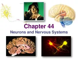
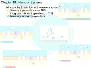
CHAPTER 28 Nervous Systems. Modules 28.1 – 28.9. NERVOUS SYSTEM STRUCTURE AND FUNCTION. 28.1 Nervous systems receive sensory input, interpret it, and send out appropriate commands. Three interconnected functions Sensory input (sensory neurons) Integration (interneurons)

E N D
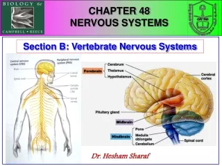
CHAPTER 28Nervous Systems Modules 28.1 – 28.9
NERVOUS SYSTEM STRUCTURE AND FUNCTION 28.1 Nervous systems receive sensory input, interpret it, and send out appropriate commands • Three interconnected functions • Sensory input (sensory neurons) • Integration (interneurons) • Motor output (motor neurons)
SENSORY INPUT INTEGRATION Sensory receptor MOTOR OUTPUT Brain and spinal cord Effector Peripheral nervoussystem (PNS) Central nervoussystem (CNS) Figure 28.1A
CNS: brain & spinal cord • PNS: nerves that carry info into and out of CNS • NS: 2 divisions
1 Sensoryreceptor 2 Sensory neuron Brain Ganglion 3 Motorneuron Spinalcord 4 Quadricepsmuscles Interneuron CNS Nerve Flexormuscles PNS Figure 28.1B
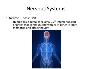
28.2 Neurons are the functional units of nervous systems • Neurons - cells transmit nervous impulses • Made of • a cell body • dendrites (highly branched fibers) • an axon (long fiber)
Myelin sheath - insulating material in vertebrates • Made of Schwann cells linked by nodes of Ranvier • Speeds up signal transmission • Multiple sclerosis (MS) involves the destruction of myelin sheaths by the immune system • Supporting cells protect, insulate, and reinforce neurons
Dendrites Signal direction Cell body Cellbody Node of Ranvier Myelin sheath Signalpathway Axon Schwann cell Nucleus Nucleus Nodes ofRanvier Schwann cell Synaptic knobs Myelin sheath Figure 28.2
NERVE SIGNALS AND THEIR TRANSMISSION 28.3 A neuron maintains a membrane potential across its membrane • Resting potential • (+) outside and (- 70 mv) inside Voltmeter Plasmamembrane Microelectrodeoutside cell –70 mV Microelectrodeinside cell Axon Neuron Figure 28.3A
28.4 A nerve signal begins as a change in the membrane potential • Stimulus changes permeability of part of plasma membrane • Ions pass through plasma membrane, changing membrane’s voltage • Causes nerve signal (electrical) to be generated
Electrical change in plasma membrane voltage (charge) from the resting potential to maximum level and back to resting potential • Action potential - nerve signal
Ions and channels involved • Sodium (Na+) outside, Potassium (K+) inside • Voltage gated (Na+) and (K+) channels where specific ions go through are found in EACH node • Change in voltage opens channels, and ions go from hi-lo concn • Sodium/Potassium pump restores resting potential
Na+ K+ Na+ K+ Additional Na+ channels open, K+ channels are closed; interior ofcell becomes more positive. 3 Na+ channels close andinactivate. K+ channelsopen, and K+ rushesout; interior of cell morenegative than outside. 4 Na+ Actionpotential 3 4 2 The K+ channels closerelatively slowly, resulting In REFRACTORY period Where the node is unable To be stimulated 5 Na+ Thresholdpotential A stimulus opens some Na+channels; if threshold is reached,action potential is triggered. 2 1 1 5 Resting potential Neuroninterior Neuroninterior Resting state: voltage gated Na+and K+ channels closed; restingpotential is maintained. 1 Return to resting state. 1 Figure 28.4
Polarized / Depolarized / Repolarized / Hyperpolarized • POLARIZED resting potential – opposite sides of membranes have opposite charges ( - inside / + outside) • DEPOLARIZED when membrane reaches threshold – 50 mV, Na+ gates open and Na+ rush in, changing inside to +++
Polarized / Depolarized / Repolarized / Hyperpolarized • REPOLARIZED Na+ gates close, K+ gates open and K+ rush out, making inside membrane (-) again • HYPERPOLARIZED K+ gates stay open longer; more K+ rush out and cell becomes – 90 mV = REFRACTORY PERIOD (node is temp unstimulatable)
HYPERPOLARIZED -> RESTING POTENTIAL • Na+ / K+ pump pumps Na+ out and K+ in to restore RP (-70 mV) • Some Na+ move to next node to change charge if reach – 50 mV, next Na+ gate opens
Na+ K+ Na+ K+ DEPOLARIZATION 3 REPLORIZATION 4 Na+ Actionpotential 3 4 2 HYPERPOLARIZATION Refractory Period 5 Na+ Thresholdpotential REACHING THRESHOLD 2 1 1 5 Resting potential Neuroninterior Neuroninterior RESTING POTENTIAL 1 Return to resting state. 1 Figure 28.4
28.5 The action potential propagates itself along the neuron Axon Action potential Axonsegment 1 Na+ Action potential K+ 2 Na+ K+ Action potential K+ 3 Na+ K+ Figure 28.5
Stronger the stimulus = more AP coming through nodes (instead of stronger AP) ex. Tap on finger versus hammer hitting finger • Action potential: all-or-none event
28.6 Neurons communicate at synapses • Synapse: • Space between two neurons or between a neuron and target (muscle) cell • Synapses - either electrical or chemical • Electrical: Action potentials pass between cells w/o neurotransmitter • Chemical: neurotransmitters cross synaptic cleft to bind to receptors on surface of the receiving cell
1 SENDINGNEURON Actionpotentialarrives Axon ofsendingneuron Vesicles Synapticknob SYNAPSE 2 3 Vesicle fuses with plasma membrane Neurotransmitteris released intosynaptic cleft SYNAPTICCLEFT 4 Receivingneuron Neuro-transmitterbinds to receptor RECEIVINGNEURON Neurotransmittermolecules Ion channels Neurotransmitter brokendown and released Neurotransmitter Receptor Ions 5 6 Ion channel opens Ion channel closes Figure 28.6
28.7 Chemical synapses make complex information processing possible • Excitatory NT: open Na+ gates for AP to move forward • Inhibitory NT: open Cl- gates = decrease next cell’s ability to develop AP • Summation: total excitation and inhibition determines whether or not next cell will move signal forward (AP go forward)
Dendrites Synaptic knobs • Neuron receiving input from 100s other neurons Myelinsheath Receivingcell body Axon Synapticknobs Figure 28.7
NERVOUS SYSTEMS 28.10 Nervous system organization usually correlates with body symmetry • Radially symmetrical animals = nerve net • Example: Hydras Nervenet Neuron A. Hydra (cnidarian) Figure 28.10A
Cephalization:concentration of nervous system in head region • Centralization: presence of CNS • Most bilaterally symmetrical animals exhibit Eye Brain Brain Brain Brain Ventralnervecord Ventralnervecord Nervecord Giantaxon Transversenerve Ganglia Segmentalganglion B. Planarian (flatworm) C. Leech (annelid) D. Insect (arthropod) E. Squid (mollusk) Figure 28.10B-E
28.11 Vertebrate nervous systems are highly centralized and cephalized CENTRAL NERVOUSSYSTEM (CNS) PERIPHERALNERVOUSSYSTEM (PNS) Brain Cranialnerve Spinal cord Spinalnerves Figure 28.11A
The brain and spinal cord contain fluid-filled spaces Dorsal rootganglion(part of PNS) Gray matter Meninges BRAIN White matter Central canal Spinal nerve(part of PNS) Ventricles Central canalof spinal cord SPINAL CORD(cross section) Spinal cord Figure 28.11B
28.12 The peripheral nervous system of vertebrates is a functional hierarchy Peripheralnervous system Sensorydivision Motordivision Sensingexternalenvironment Sensinginternalenvironment Autonomicnervous system(involuntary) Somaticnervous system(voluntary) Sympatheticdivision Parasympatheticdivision Figure 28.12A
Autonomic nervous system: involuntary control over the internal organs • Somatic nervous system: voluntary control over skeletal muscles • Motor division of the PNS
28.13 Opposing actions of sympathetic and parasympathetic neurons regulate the internal environment • Autonomic NS: • Parasympathetic division: resting / digesting • Sympathetic division: fight / flight
PARASYMPATHETIC DIVISION SYMPATHETIC DIVISION Eye Brain Constrictspupil Dilatespupil Salivaryglands Stimulatessalivaproduction Inhibitssalivaproduction Lung Relaxesbronchi Constrictsbronchi Acceleratesheart Slowsheart Adrenalgland Heart Stimulatesepinephrineand norepi-nephrine release Liver Spinalcord Stomach Stimulatesstomach,pancreas,and intestines Stimulatesglucoserelease Pancreas Inhibitsstomach,pancreas,and intestines Intestines Bladder Stimulatesurination Inhibitsurination Figure 28.13
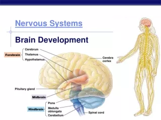
Cerebrum Forebrain Thalamus Cerebralcortex Hypothalamus Pituitary gland Midbrain Pons Medullaoblongata Hindbrain Spinal cord Cerebellum Figure 28.15A
Hemisphere – left/right Left cerebralhemisphere Right cerebralhemisphere Corpuscallosum Basalganglia Figure 28.15B
28.16 Parts of CNS / Brain • Cerebral cortex: voluntary motion; higher function (memory and creativity); conscious mind; speech; problem solving; sensations – smell, sights, temperature, etc • Olfactory lobe: smell • Thalamus: relay sensory input and distribute to appropriate part of cerebral cortex • Hypothalamus: body homeostasis – hormone level, temperature, hunger, thirst, pain, etc
Pons: relay center cerebellum • Medulla (oblongata): vital functions – breathing, heart rate, CO2 level • Cerebellum: muscle movement and balance • Spinal cord: brain to body – reflex arc
CHAPTER 29The Senses Modules 29.1 – 29.3
29.1 Sensory inputs become sensations and perceptions in the brain • Sensation • Awareness of sensory stimuli • Perception • Brain’s full integration of sensory data Figure 29.1
SENSORY RECEPTION 29.2 Sensory receptor cells convert stimuli into electrical energy • Sensory receptors (sight, touch, sound, smell, taste)
Sugarmolecule Sugar molecule Taste pore Ion Tongue Ionchannels Taste bud Sensoryreceptorcells Receptor cellmembrane 2 3 Sugarbinding Receptorpotential Sensory neuron Neuro-transmittermolecules Sensoryreceptorcell 1 Taste bud anatomy Sensoryneuron Action potential 4 Synapse mV No sugar Sugar present 5 Action potentials Figure 29.2A
Brain distinguishes different types of stimuli • Action potentials transmitted to CNS via sensory neurons
Interneurons Sugarreceptor Salt receptor BRAIN TASTEBUD Sensoryneurons No sugar No salt Increasing sweetness Increasing saltiness Figure 29.2B
29.3 Specialized sensory receptors detect five categories of stimuli • Pain receptors • Sense dangerous stimuli • Thermoreceptors • Detect heat or cold • Mechanoreceptors • Respond to mechanical energy (touch, pressure, and sound)
Heat Lighttouch Pain Cold (Hair) Lighttouch Epidermis Dermis Nerve Touch Strongpressure Figure 29.3A
Stretch receptors and hair cells are two types of mechanoreceptors “Hairs” ofreceptor cell Moreneurotransmitter Neurotransmitterat synapse Lessneurotransmitter Sensoryneuron Actionpotentials (1) Receptor cell at rest (2) Fluid moving in one direction (3) Fluid moving in other direction Figure 29.3B
Eye Infraredreceptor • Respond to electricity, magnetism, and light • Photoreceptors sense light • They are the most common electromagnetic receptors • Electromagnetic receptors Figure 29.3D
29.5 Vertebrates have single-lens eyes Sclera Choroid Retina Muscle Ligament Fovea(center ofvisual field) Cornea Iris Opticnerve Pupil Aqueoushumor Lens Arteryand vein Vitreoushumor Blind spot Figure 29.5
Cornea and lens focus light on photoreceptor cells in the retina • Photoreceptors most concentrated in fovea • two eyes compensates for blind spot • blind spot optic nerve passes through retina • Human eye
Muscle contracted Muscle relaxed Choroid Ligaments Retina Light from anear object Light from adistant object Lens NEAR VISION(ACCOMMODATION) DISTANCE VISION Figure 29.6
29.8 Our photoreceptor cells are rods and cones • photoreceptor cells • Rods (contrast – black/white) • Cones (color) ROD Cell body CONE Synapticknobs Membranous discscontaining visual pigments Figure 29.8A
Retina Photoreceptors Neurons Cone Rod Opticnervefibers Retina Fovea Opticnerve Figure 29.8B