Download
1 / 12
120 likes | 280 Views
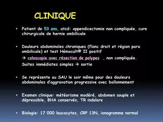
RTC 3D ORGANE SITUATION CLINIQUE. Cas Clinique. Données cliniques. CONTOURAGE. Coupes TDM avec volumes cibles. CONTOURAGE. Coupes TDM avec volumes cibles. CONTOURAGE. Coupes TDM avec volumes cibles. DOSIMETRIE. Volumes cible : Points d’intérêt Axe des faisceaux pelviens =

E N D
RTC 3DORGANESITUATION CLINIQUE Cas Clinique
CONTOURAGE • Coupes TDM avec volumes cibles
CONTOURAGE • Coupes TDM avec volumes cibles
CONTOURAGE • Coupes TDM avec volumes cibles
DOSIMETRIE • Volumes cible : • Points d’intérêt • Axe des faisceaux pelviens = • Axes des faisceaux réduits = a • Faisceaux pelviens • Axe = • Orientation = • Pondération = • Caches = mm autour du PTV1 • Faisceaux réduits : • Axe = axe réduit • Orientation = selon scanner dosimétrique • Pondération = selon scanner dosimétrique • Caches = mm autour du PTV2
DOSIMETRIE • Image des faisceaux
DOSIMETRIE • Image des faisceaux