Download
1 / 25
250 likes | 273 Views
Depolarization. Initially, this is a local electrical event called end plate potential Later, it ignites an action potential that spreads in all directions across the sarcolemma. Action Potential: Electrical Conditions of a Polarized Sarcolemma.

E N D
Depolarization • Initially, this is a local electrical event called end plate potential • Later, it ignites an action potential that spreads in all directions across the sarcolemma
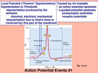
Action Potential: Electrical Conditions of a Polarized Sarcolemma • The outside (extracellular) face is positive, while the inside face is negative • This difference in charge is the resting membrane potential Figure 9.8a
Action Potential: Electrical Conditions of a Polarized Sarcolemma • The predominant extracellular ion is Na+ • The predominant intracellular ion is K+ • The sarcolemma is relatively impermeable to both ions Figure 9.8a
Action Potential: Depolarization and Generation of the Action Potential • An axonal terminal of a motor neuron releases ACh and causes a patch of the sarcolemma to become permeable to Na+ (sodium channels open) Figure 9.8b
Action Potential: Depolarization and Generation of the Action Potential • Na+ enters the cell, and the resting potential is decreased (depolarization occurs) • If the stimulus is strong enough, an action potential is initiated Figure 9.8b
Action Potential: Propagation of the Action Potential • Polarity reversal of the initial patch of sarcolemma changes the permeability of the adjacent patch • Voltage-regulated Na+ channels now open in the adjacent patch causing it to depolarize Figure 9.8c
Action Potential: Propagation of the Action Potential • Thus, the action potential travels rapidly along the sarcolemma • Once initiated, the action potential is unstoppable, and ultimately results in the contraction of a muscle Figure 9.8c
Action Potential: Repolarization • Immediately after the depolarization wave passes, the sarcolemma permeability changes • Na+ channels close and K+ channels open • K+ diffuses from the cell, restoring the electrical polarity of the sarcolemma Figure 9.8d
Action Potential: Repolarization • Repolarization occurs in the same direction as depolarization, and must occur before the muscle can be stimulated again (refractory period) • The ionic concentration of the resting state is restored by the Na+-K+ pump Figure 9.8d
Excitation-Contraction Coupling • Once generated, the action potential: • Is propagated along the sarcolemma • Travels down the T tubules • Triggers Ca2+ release from terminal cisternae • Ca2+ binds to troponin and causes: • The blocking action of tropomyosin to cease • Actin active binding sites to be exposed
Excitation-Contraction Coupling • Myosin cross bridges alternately attach and detach • Thin filaments move toward the center of the sarcomere • Hydrolysis of ATP powers this cycling process • Ca2+ is removed into the SR, tropomyosin blockage is restored, and the muscle fiber relaxes
Role of Ionic Calcium (Ca2+) in the Contraction Mechanism • At low intracellular Ca2+ concentration: • Tropomyosin blocks the binding sites on actin • Myosin cross bridges cannot attach to binding sites on actin • The relaxed state of the muscle is enforced • At higher intracellular Ca2+ concentrations: • Additional calcium binds to troponin (inactive troponin binds two Ca2+) • Calcium-activated troponin binds an additional two Ca2+ at a separate regulatory site Figure 9.11a
Sequential Events of Contraction • Cross bridge formation – myosin cross bridge attaches to actin filament • Working (power) stroke – myosin head pivots and pulls actin filament toward M line • Cross bridge detachment – ATP attaches to myosin head and the cross bridge detaches • “Cocking” of the myosin head – energy from hydrolysis of ATP cocks the myosin head into the high-energy state
ADP Myosin head (high-energy configuration) Pi 1 Myosin head attaches to the actin myofilament, forming a cross bridge. Thin filament ADP ADP ATP Thick filament hydrolysis Pi 2 Inorganic phosphate (Pi) generated in the previous contraction cycle is released, initiating the power (working) stroke. The myosin head pivots and bends as it pulls on the actin filament, sliding it toward the M line. Then ADP is released. 4 As ATP is split into ADP and Pi, the myosin head is energized (cocked into the high-energy conformation). ATP Myosin head (low-energy configuration) ATP 3 As new ATP attaches to the myosin head, the link between myosin and actin weakens, and the cross bridge detaches. Figure 9.12
Contraction of Skeletal Muscle (Organ Level) • Contraction of muscle fibers (cells) and muscles (organs) is similar • The two types of muscle contractions are: • Isometric contraction – increasing muscle tension (muscle does not shorten during contraction) • Isotonic contraction – decreasing muscle length (muscle shortens during contraction)
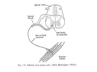
Motor Unit: The Nerve-Muscle Functional Unit • A motor unit is a motor neuron and all the muscle fibers it supplies • The number of muscle fibers per motor unit can vary from four to several hundred • Muscles that control fine movements (fingers, eyes) have small motor units
Motor Unit: The Nerve-Muscle Functional Unit • Large weight-bearing muscles (thighs, hips) have large motor units • Muscle fibers from a motor unit are spread throughout the muscle; therefore, contraction of a single motor unit causes weak contraction of the entire muscle
Muscle Twitch • A muscle twitch is the response of a muscle to a single, brief threshold stimulus • There are three phases to a muscle twitch • Latent period • Period of contraction • Period of relaxation
Phases of a Muscle Twitch • Latent period – first few msec after stimulus; EC coupling taking place • Period of contraction – cross bridges from; muscle shortens • Period of relaxation – Ca2+ reabsorbed; muscle tension goes to zero Figure 9.14a
Graded Muscle Responses • Graded muscle responses are: • Variations in the degree of muscle contraction • Required for proper control of skeletal movement • Responses are graded by: • Changing the frequency of stimulation • Changing the strength of the stimulus
Muscle Response to Varying Stimuli • A single stimulus results in a single contractile response – a muscle twitch • Frequently delivered stimuli (muscle does not have time to completely relax) increases contractile force – wave summation Figure 9.15
Muscle Response to Varying Stimuli • More rapidly delivered stimuli result in incomplete tetanus • If stimuli are given quickly enough, complete tetanus results Figure 9.15
Muscle Response: Stimulation Strength • Threshold stimulus – the stimulus strength at which the first observable muscle contraction occurs • Beyond threshold, muscle contracts more vigorously as stimulus strength is increased • Force of contraction is precisely controlled by multiple motor unit summation • This phenomenon, called recruitment, brings more and more muscle fibers into play
Treppe: The Staircase Effect • Staircase – increased contraction in response to multiple stimuli of the same strength • Contractions increase because: • There is increasing availability of Ca2+ in the sarcoplasm • Muscle enzyme systems become more efficient because heat is increased as muscle contracts
Treppe: The Staircase Effect Figure 9.18