Download
1 / 73
870 likes | 1.52k Views
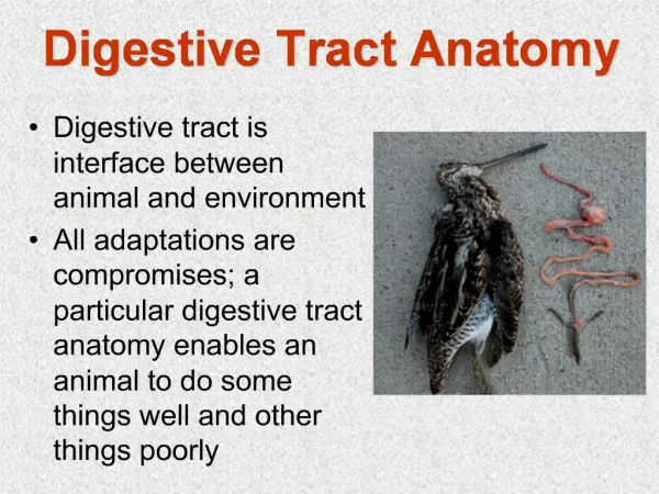
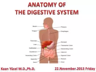
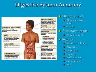
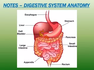
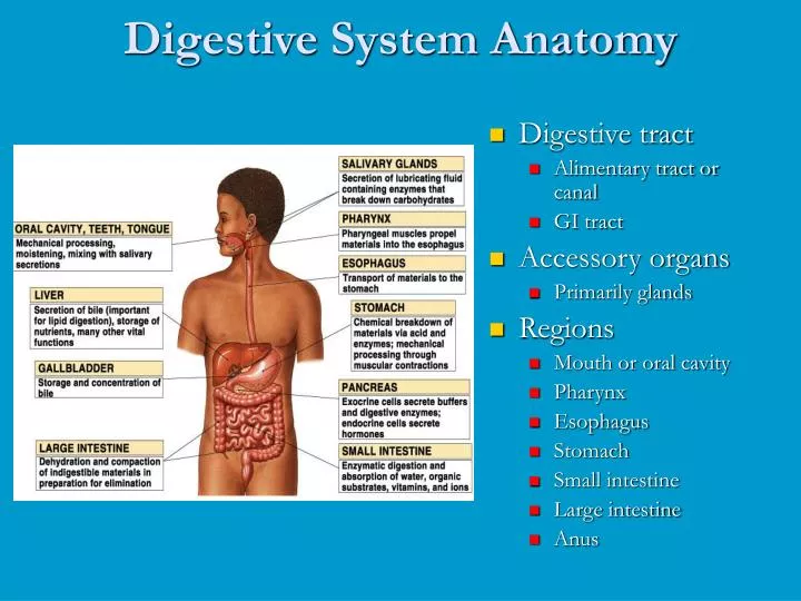
Digestive System Anatomy. Digestive tract Alimentary tract or canal GI tract Accessory organs Primarily glands Regions Mouth or oral cavity Pharynx Esophagus Stomach Small intestine Large intestine Anus. Digestive Tract Histology. Nervous regulation Involves enteric nervous system

E N D
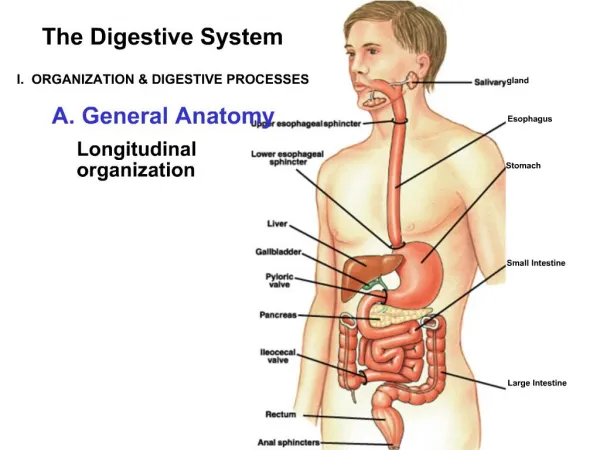
Digestive System Anatomy • Digestive tract • Alimentary tract or canal • GI tract • Accessory organs • Primarily glands • Regions • Mouth or oral cavity • Pharynx • Esophagus • Stomach • Small intestine • Large intestine • Anus
Nervous regulation Involves enteric nervous system Types of neurons: sensory, motor, interneurons Coordinates peristalsis and regulates local reflexes Chemical regulation Production of hormones Gastrin, secretin Production of paracrine chemicals Histamine Help local reflexes in ENS control digestive environments as pH levels Digestive System Regulation
Peritoneum and Mesenteries • Peritoneum • Visceral: Covers organs • Parietal: Covers interior surface of body wall • Retroperitoneal: Behind peritoneum as kidneys, pancreas, duodenum • Mesenteries • Routes which vessels and nerves pass from body wall to organs • Greater omentum • Lesser omentum
Oral Cavity • Mouth or oral cavity • Vestibule: Space between lips or cheeks and alveolar processes • Oral cavity proper • Lips (labia) and cheeks • Palate: Oral cavity roof • Hard and soft • Palatine tonsils • Tongue: Involved in speech, taste, mastication, swallowing
Teeth • Two sets • Primary, deciduous, milk: Childhood • Permanent or secondary: Adult (32) • Types • Incisors, canine, premolar and molars
Salivary Glands • Produce saliva • Prevents bacterial infection • Lubrication • Contains salivary amylase • Breaks down starch • Three pairs • Parotid: Largest • Submandibular • Sublingual: Smallest
Pharynx Nasopharynx Oropharynx: Transmits food normally Laryngopharynx: Transmits food normally Esophagus Transports food from pharynx to stomach Passes through esophageal hiatus (opening) of diaphragm and ends at stomach Hiatal hernia Sphincters Upper Lower Pharynx and Esophagus
Deglutition (Swallowing) • Three phases • Voluntary • Bolus of food moved by tongue from oral cavity to pharynx • Pharyngeal Reflex: Upper esophageal sphincter relaxes, elevated pharynx opens the esophagus, food pushed into esophagus • Esophageal • Reflex: Epiglottis is tipped posteriorly, larynx elevated to prevent food from passing into larynx
Functions • Ingestion: Introduction of food into stomach • Mastication: Chewing • Propulsion • Deglutition: Swallowing • Peristalsis: Moves material through digestive tract
Stomach Anatomy: • Openings • Gastroesophageal: To esophagus • Pyloric: To duodenum • Regions • Cardiac • Fundus • Body • Pyloric
Stomach Histology: • Layers • Serosa or visceral peritoneum: Outermost • Muscularis: Three layers • Outer longitudinal • Middle circular • Inner oblique • Submucosa • Mucosa
Stomach Histology • Rugae: Folds in stomach when empty • Gastric pits: Openings for gastric glands • Contain cells • Surface mucous: Mucus • Mucous neck: Mucus • Parietal: Hydrochloric acid and intrinsic factor • Chief: Pepsinogen • Endocrine: Regulatory hormones
Small Intestine • Site of greatest amount of digestion and absorption • Divisions • Duodenum • Jejunum • Ileum: Peyer’s patches or lymph nodules • Modifications • Circular folds or plicae circulares, villi, lacteal, microvilli • Cells of mucosa • Absorptive, goblet, granular, endocrine
Small Intestine Secretions • Mucus • Protects against digestive enzymes and stomach acids • Digestive enzymes • Disaccharidases: Break down disaccharides to monosaccharides • Peptidases: Hydrolyze peptide bonds • Nucleases: Break down nucleic acids • Duodenal glands • Stimulated by vagus nerve, secretin, chemical or tactile irritation of duodenal mucosa
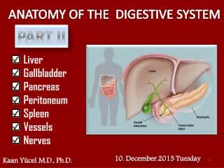
Liver • Lobes • Major: Left and right • Minor: Caudate and quadrate • Ducts • Common hepatic • Cystic • From gallbladder • Common bile • Joins pancreatic duct at hepatopancreatic ampulla
Functions of the Liver • Bile production • Salts emulsify fats, contain pigments as bilirubin • Storage • Glycogen, fat, vitamins, copper and iron • Nutrient interconversion • Detoxification • Hepatocytes remove ammonia and convert to urea • Phagocytosis • Kupffer cells phagocytize worn-out and dying red and white blood cells, some bacteria • Synthesis • Albumins, fibrinogen, globulins, heparin, clotting factors
Gallbladder • Bile is stored and concentrated • Stimulated by cholecystokinin and vegal stimulation • Dumps into small intestine • Production of gallstones possible • Drastic dieting with rapid weight loss
Anatomy Endocrine Pancreatic islets produce insulin and glucagon Exocrine Acini produce digestive enzymes Regions: Head, body, tail Secretions Pancreatic juice (exocrine) Trypsin Chymotrypsin Carboxypeptidase Pancreatic amylase Pancreatic lipases Enzymes that reduce DNA and ribonucleic acid Pancreas
Movement in small intestine: • Mixing: Segmental contraction that occurs in small intestine • Secretion: Lubricate, liquefy, digest • Digestion: Mechanical and chemical • Absorption: Movement from tract into circulation or lymph • Elimination: Waste products removed from body
Large Intestine: • Extends from ileocecal junction to anus • Consists of cecum, colon, rectum, anal canal • Movements sluggish (18-24 hours)
Large Intestine • Cecum • Blind sac, vermiform appendix attached • Colon • Ascending, transverse, descending, sigmoid • Rectum • Straight muscular tube • Anal canal • Internal anal sphincter (smooth muscle) • External anal sphincter (skeletal muscle) • Hemorrhoids: Vein enlargement or inflammation
Secretions of Large Intestine • Mucus provides protection • Parasympathetic stimulation increases rate of goblet cell secretion • Pumps • Exchange of bicarbonate ions for chloride ions • Exchange of sodium ions for hydrogen ions • Bacterial actions produce gases called flatus
Movement in Large Intestine • Mass movements • Common after meals • Local reflexes in enteric plexus • Gastrocolic: Initiated by stomach • Duodenocolic: Initiated by duodenum • Defecation reflex • Distension of the rectal wall by feces • Defecation • Usually accompanied by voluntary movements to expel feces through abdominal cavity pressure caused by inspiration