Download

1 / 82
1.05k likes | 2.29k Views
Digestive System Digestive Glands. Components of Digestive Glands. ◇ small digestive glands: found in the wall of digestive tract. ◇ accessory glands (large digestive glands):. salivary glands pancreas liver. General Structure of Digestive Glands.

E N D
Digestive System Digestive Glands
Components of Digestive Glands ◇small digestive glands: found in the wall of digestive tract. ◇accessory glands (large digestive glands): salivary glands pancreas liver
General Structure of Digestive Glands ◇Parenchyma: ( functional portion of an organ ) acini / gland cells ducts ◇Stroma: ( non-functional portion of an organ ) capsule CT inside the organ.
Salivary Glands General structure of the large salivary glands serous acinus mucous acinus seromucous / mixed acinus acinus types of acinus Parenchyma intercalated duct striated/secretory duct interlobular duct excretory duct duct
serous acinus seromucous / mixed acinus mucous acinus intercalated duct striated /secretory duct demilume Model ( structure of the large salivary glands)
seromucous /mixed acinus mucous acinus serous acinus Model Section (H&E)
Salivary Glands Structural Characteristic of gland Cell: serous acinus:comprised by serous cells. *zymogen granules in apical cytoplasm. mucous acinus:comprised by serous cells. *mucinogen granules in cytoplasm. seromucous acinus:comprised by both cells. / mixed acinus*demilume
serous acinus: *zymogen granules in apical cytoplasm.
Serous cell: basal lamina (bl); connective tissue (ct); desmosome (d); endothelium (en); Golgi complex (g); intercellular space (is); lumen (l); microvilli (mi); mitochondria (m); nucleus (nu); RER (re); secretion granule (sg). 10.000x.
mucous acinus: *mucinogen granules in cytoplasm. n
Detail of mucous cell: Sero-mucous cell (sc); Golgi complex (gc); intercellular space (is); secretion granule (sg); lumen (l); nucleus (n); RER (re). 10000x. sg is re n
seromucous acinus / mixed acinus demilume
Salivary Glands acinus duct intercalated duct: simple squamous/cuboidal epith. striated/secretory duct: simple tall columnar epith. interlobular duct: pseudostratified columnar epith. excretory duct: stratified squamous epith.
Salivary Glands intercalated duct: simple squamous or cuboidal epith.
Salivary Glands intercalated duct: simple squamous or cuboidal epith.
LM EM
Detail ofintercalated duct cell basal lamina (bl); desmosome (d); nerve fibers (nf); Golgi complex (g); intercellular space (is); mitochondria (m); nucleus (nu); RER (re). 13.000x. g m re nu bl EM
Salivary Glands striated/secretory duct:simple tall columnar epith.
Salivary Glands striated/secretory duct Note: the basal striations
Striated duct: blood vessel (bv); connective tissue (ct); lumen (l); nucleus (nu). ×2750.
Basal portion of striated duct cell: basal lamina (bl); basal membrane pleat (bmp); collagen fiber (cf); mitochondria (m); nucleus (nu). ×16.500 .
Salivary Glands striated/secretory duct The secretory ducts,which are continuous with the intercalated ducts,are wider and lined with a simple columnar epithelium. As the secretion from the acini passes through the secretory ducts,the epithelium can re-absorb sodium(Na+) and water from the lumen to the interstitium(间质)and transport potassium (K+) into the saliva,thus changing the consistency of the saliva. The secretory ducts drain into interlobular ducts which run between lobules.
Salivary Glands interlobular duct: pseudostratified columnar epith. excretory duct: stratified squamous epith.
Salivary glands include *Parotid G. *Submandibular G. * Sublingual G. Function: * moistening food. * carbohydrate digestion. * Secrete IgA.
Structural Features of Salivary Glands Parotid G. Submandibular G. Sublingual G. Serous A. +++ +++++ + Mucous A. none + +++ Mixed A. none + + Inter- calated D none short long few long short Striated D
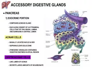
Pancreas The pancreas is a lobular organ. The pancreas has both exocrine functions (releases digestive enzyme secretions into the intestines) and endocrine functions (releases hormones into the blood).
Pancreas Parenchyma endocrine gland exocrine gland produces hormones produces pancreatic juice
Parenchyma make section
Parenchyma endocrine gland endocrine gland exocrine gland exocrine gland exocrine gland
Parenchyma exocrine gland endocrine gland
Pancreas Exocrine Pancreas Acini Ducts intercalated ducts interlobular ducts main pancreatic ducts • wholly consists of serous acini. • small centroacinar cells in the lumen. major duodenal papilla
Exocrine Pancreas Here is another look at the Exocrine Pancreas and the Acini (white dotted lines) which make it up. Central Acinar Cells The yellow arrows are pointing to the characteristic centroacinar cells which are the key to identifying the pancreas. Notice how they stand out against the dark cells of the acini. Pancreas Acinus
Exocrine Pancreas Centroacinar Cell Pancreas Acinus Acinar Cell Low power (LM) high power (LM)
Exocrine Pancreas • Ducts • Intercalated ducts: simple squamous • or cuboidal epith. • Interlobular ducts:columnar epith. • pancreatic ducts: tall columanr epth. (similar to salivary glands. REMEMBER: there are no striated ducts in the pancreas. ) major duodenal papilla
Exocrine Pancreas It releases the pancreatic juice. pancreatic juice contain many kinds of enzyme: * amylase(胰淀粉酶):hydrolyses starch & glycogen . * lipase(胰脂肪酶):hydrolyzes triglycerides(甘油三酯). into fattyacids and mono-glycerides(甘油单酯). * cholesterol esterase(胆固醇酯酶) :breaks down cholesterol esters(酯)into cholesterol and a fatty acid. * trypsin( 胰蛋白酶)and chymotrypsin(糜蛋白酶) : hydrolyze proteins. * ribonuclease(核糖核酸酶)& deoxyribonuclease(脱氧核 糖核酸酶):split nucleic acids.
Pancreas Endocrine Pancreas • Islets of Langerhansscatter throughout the exocrine pancreas. • Three types of cells : • A-cells(20%) glucagon • B-cells(75%) insulin • D-cells( 5% ) somatostatin • Capillaries:each islet is richly supplied with blood vessels.
Pancreas Endocrine Pancreas D-cells A-cells B-cells A-cells A-cells B-cells
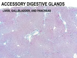
Liver General Introduction *The largest gland (~ 2% of body weight in adult). * Respectively receives both venous & arterial blood through the portal V. (~75%) & hepatic A (~25%). * CT of capsule extended into the parenchyma, forming “classical” liver loblules. * Functions as an exocrine gland (secreting bile) and other very important roles.
Liver liver loblules section
Liver Structures of Liver Lobule
Liver Structures of Liver Lobule
Liver Structures of Liver Lobule *six-sided prism with a central V. at its center. *sheets of hepatocytes( orhepatic plates) extend radially from the central V. * sinusoids between hepatic plates. portal triads ( or portal area): in the corner of lobules.
Liver Structures of Liver Lobule hepatic plates
Liver Portal Triads (or Portal Area) * Definition: The area of CT found in the angles where adjacent hepatic lobules meet. * Components: CT. + portal triad interlobularA interlobularV interlobularbile duct
Liver Portal Triads (or Portal Area)