Download
1 / 12
271 likes | 870 Views
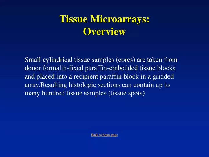
Tissue Microarrays: Overview. Small cylindrical tissue samples (cores) are taken from donor formalin-fixed paraffin-embedded tissue blocks and placed into a recipient paraffin block in a gridded array.Resulting histologic sections can contain up to many hundred tissue samples (tissue spots).

E N D
Tissue Microarrays:Overview Small cylindrical tissue samples (cores) are taken from donor formalin-fixed paraffin-embedded tissue blocks and placed into a recipient paraffin block in a gridded array.Resulting histologic sections can contain up to many hundred tissue samples (tissue spots) Back to home page
Beecher Instruments:Manual Tissue Microarray Apparatus Needle holder: slides up & down to collect & deposit tissue cores Calipers: move the needle holder in precise increments in the x & y directions to form the TMA grid Hollow needles: punch holes in donor & recipient blocks Holds recipient block steady during TMA manufacture Stage to support donor block during tissue sampling Back to home page
Review of TMA manufacture A hole is made into the recipient block at a predetermined position Back to home page
Review of TMA manufacture The stage is placed over the recipient block. A donor block is placed on the stage. The guide slide is positioned on the block to best match the contours of the tissue. Note that the guide slide has been circled to indicate the position of the target tissue. Back to home page
Review of TMA manufacture With the guide slide placed in the best approximation with the tissue in the block, the donor block is moved such that the circled area of target tissue is immediately under the needle Back to home page
Review of TMA manufacture Note that the maker of the TMA is guided solely by the gross visual examination of tissue contours and the circled area. No microscopic assessments are made during this phase of TMA manufacture. Back to home page
Review of TMA manufacture The slide is removed, and the needle is driven into the donor block. Back to home page
Review of TMA manufacture The needle, containing the tissue sample, is withdrawn from the donor block, and the stage and the donor block are removed. The tissue-containing needle is lowered over the previously-made hole in the recipient block. Back to home page
Review of TMA manufacture The tissue core is inserted into the recipient block by pressing on the stylus. Back to home page
Tissue core in recipient block Back to home page
The tissue cores sizes, their spacing, and their arrangement in the block are at the discretion of the designer of the TMA. Back to home page
Increasing magnification of a TMA Back to home page