Download
1 / 28
280 likes | 502 Views
Microarrays at WMRGL. By Nicola James Genetic Technologist Day 02.06.08. Overview. Introduction Arrays in our department Diagnostic Array CGH: BlueGnome CytoChips Research SNP Arrays: Affymetrix Protocol breakdown Result Validation Case examples. Resolution Scale.

E N D
Microarrays at WMRGL By Nicola James Genetic Technologist Day 02.06.08
Overview • Introduction • Arrays in our department Diagnostic Array CGH: BlueGnome CytoChips Research SNP Arrays: Affymetrix • Protocol breakdown • Result Validation • Case examples
Resolution Scale Increased size of imbalance 1bp 6Kb 5Mb >10Mb Sequencing DNA Microarrays Cytogenetics Increased resolution
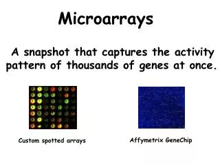
Array CGH Ratio Test Genomic DNA Reference Genomic DNA Cot-1 DNA Genomic clones or oligos spotted on glass slide DNA gain DNA loss Position on Sequence
Array Platforms • Diagnostic: 1Mb CytoChip Duplicate sub arrays (dye swap) Extensive replicates of backbone and disease specific clones (90 genetic conditions) >4000 clones cover whole genome Median resolution 565Kb • Research: Affymetrix arrays Detect SNPs Mean genomic spacing 250Kb Pt Ref Pt Ref
Disease Specific Clones www.cytochip.com/
Array CGH Protocol Referral received from Clinical Genetics Report Blood cultured and DNA extracted FISH/molecular validation studies DNA quality assessment Result interpretation and analysis Random prime labelling of DNA Image acquisition via laser scanning Labelled DNA purification and assessment Automated slide washing/drying Co-precipitation with Cot-1 Overnight hybridisation to array
DNA Quality Control • DNA quality assessed using Nanodrop spectrophotometer and gel electrophoresis. • Crude band assessment on gel. • High molecular weight DNA required. • Degraded DNA appears as a grey smear (smaller fragments migrate further through gel).
DNA Optical Density • NanoDrop measures light absorbancy ratios. • DNA absorbs at 260nm, protein contaminants at 280nm, organic compounds at 230nm • Ideally: Concentration >100ng/µl 260/280 1.8>2.2 260/230 1.8>2.2
DNA Labelling • DNA labelled with Cy3 and Cy5 (reference DNA also) • Reactive water soluble fluorescent dyes with side groups so that they can chemically link to nucleic acids. Cy3 excited at 550nm emits at 570nm (red part of spectrum) Cy5 excited at 649nm emits at 670nm (far red part of spectrum) • Scanner can easily distinguish between the two: Cy3 (although red in colour) appears green on array Cy5 (although blue/green in colour) appears red on array
Random Prime Labelling Reaction Reaction mix contains high molecular weight genomic DNA, random primers, Cy-dCTPs, dNTPs (lower concentration Cs) Catalysed by Klenow enzyme Genomic DNA Extension by Klenow Overnight @ 37oC Primer Annealing Heat Denaturation Snap Chill Addition of EDTA the following morning stops the reaction Labelled DNA needs quantification and purification
Purification + Dye Incorporation Spin 2000g 1min Labelled DNA Nanodrop QC: Concentration >150ng/µl Dye incorporation >3pmol/µl Measure dye incorporation on Nanodrop
Pre-Hybridisation • Test and reference combined Test Cy3 + Reference Cy5 Test Cy5 + Reference Cy3 • Precipitation with Na acetate and ethanol • Pellet resuspended in hybridisation solution: Cot 1 DNA - blocks repetitive sequences Herring sperm DNA - minimises non-specific binding Formamide lowers denaturation temp to 75°C Dextran sulphate - ensures labelled DNA is close to the array surface • Denaturation at 75°C • Prehybridisation at 37°C for 1 hour
Hybridisation • Barcoded glass array slides (cytochips) clamped in place. • Hybridisation program selected. • Samples injected into appropriate chamber. • Automated hybridisation for 21 hours. • Automated slide washing/drying. Tecan HS 4800 Pro Hyb Station
Scanning • Array slides removed from hyb station. • Slides protected in cytochip holder – slot into scanner. • Agilent Scanner – takes up to 48 cytochips. • Laser scanner – simultaneously scans red and green signals. • Rapid – approx 10 mins per whole array.
Scanning data interpreted by BlueFuse analytical software. Analysis Process: 1. Raw data input 2. Grid alignment and quantification 3. Normalisation and exclusion 4. Dye swap integration 5. Chromosome analysis 6. Database analysis Analysis format tabular and diagrammatical – links to Ensembl. Analysis
Validation Techniques • FISH Using RPCI BAC clones – labelled in house Quick, relatively cheap, section within department already Positional information obtained for probands and parents • Molecular Assays (MLPA) Validation of smaller imbalances (<1Mb) • Alternative array platform Affymetrix SNP arrays X array?
Case Example 1 7 year old female Developmental and speech delay Mild dysmorphism Behavioural problems ?1p36 deletion
Case Example 7 BACs deleted on 2q Approx 5.9~7.8Mb in size ? de novo FISH validation required
FISH Validation Mother Proband Father Both parents normal (position and copy number OK) Deletion de novo in proband
Case Example 2 • Originally referred in 1991 • FTT and DevDelay • Cytogenetics reported inversion on chromosome 16 • Array CGH identified a 7 BAC deletion (5-6Mb, 65 HGNC mapped genes) • Reconfirmed by cytogenetics and FISH as a deletion rather than an inversion • Breakpoint close to CDH1 gene involved in gastric cancer • MPLA confirmed CDH1 region not involved
Case Example 3 low set ears corneal opacites syndactyly ?18q deletion narrow external auditory meatus (tube running from outer to inner ear)
Case Example 3 • 8 BAC deletion • 7q21.2 to 7q21.3 • 6.4 to 6.75Mb • G band visible but at limits of cytogenetic resolution • Mother normal • Paternal sample not available
1.2Mb critical region SHFM1 Locus Characters include: Syndactyly Median clefts of hands and feet Learning difficulties Cleft palate Explains some of the phenotypic features of patient
Summary • Array CGH is a high resolution technique, but time consuming and relatively expensive. • Validation required to confirm results. • Balanced rearrangements not detected. • Distinguishing between rare polymorphisms and genuine pathogenic imbalances difficult. • Anticipated new version CytoChip with more replicates will eliminate the need for dye swap. • Robotic labelling?! • Other arrays: Affymetrix SNP projects in lab (currently non-diagnostic use).
Acknowledgements Dominic McMullan Judith Walker Lisa Cooper-Charles Lee Silcock