Download

1 / 17
1.67k likes | 8.37k Views
Practical Hematology Lab. - LAB 5 -. Osmotic Fragility Test. Definition. The osmotic fragility test is a measure of the ability of the red cells to take up fluid without lysing.

E N D
Practical Hematology Lab - LAB 5 - Osmotic Fragility Test
Definition • The osmotic fragility test is a measure of the ability of the red cells to take up fluid without lysing. • Or it is a test to measures red blood cell (RBC) resistance to hemolysis when exposed to a series of increasingly dilute saline solutions. Factors Affecting On The Osmotic Fragility • The primary factor affecting the osmotic fragility test is the shape of the red cell, which, in turn, depends on the • volume • surface area • functional state of the red blood cell membrane.
1. Increased Surface – To – Volume Ratios • it is more resistant to hemolysis and has decreased fragility • The larger the amount of red cell membrane (surface area) in relation to the size of the cell, the more fluid the cell is capable of absorbing before rupturing . As • Iron-deficiency anemia • Thalassemia • Sickle cell anemia • Liver disease and any condition associated with the presence of target cells
The target cell has the largest surface area (amount of membrane) for its size and therefore shows decreased fragility. • As the red cell takes in fluid it becomes more round (spherocytic). It therefore follows that the spherocyte has the smallest surface area for its volume, ruptures the most quickly, and has increased fragility.
2. Decreased Surface –To– Volume Ratios Increased osmotic fragility (decreased resistance) is found in hemolytic anemias hereditary spherocytosis And whenever spherocytes are found.
3- Functional state of the cell membrane • The aged erythrocytes will be hemolyzedeasily, hence the more healthy cell membrane the less fragile cells such as • Reticulocytes • Splenectomy • Polycythemia
Specimen • Heparinized venous blood , or, 15 to 20 mL of defibrinated whole blood. • The test should be set up within 2 hours of collection, or within 6 hours if the blood is refrigerated.
Principle • In the osmotic fragility test, whole blood is added to varying concentrations of buffered sodium chloride solution and allowed to incubate at room temperature. • The amount of hemolysis in each saline concentration is then determined by reading the supernatants on a spectrophotometer.
Procedure Prepare dilutions of buffered sodium chloride and place in the appropriately labeled test tube (Table).
Procedure Mix the preceding dilutions well, using Parafilm to cover each test tube while mixing. Transfer 5 mL of each dilution to a second set of test tubes labeled # 1 through #14. Add 0.05 mL of the patient's heparinized blood to each of the 14 test tubes. Repeat, adding the normal control blood to the set of 14 control test tubes. Mix each test tube immediately by gentle inversion. Allow the test tubes to stand at room temperature for 30 minutes.
Procedure • Remix the test tubes gently and centrifuge at 1200 to 1500 g for 5 minutes. • Carefully transfer the supernatants to cuvettes and read on a spectrophotometer at a wavelength of 540 nm. Set the optical density at 0, using the supernatant in test tube #1, which represents the blank, or 0% hemolysis. Test tube #14 represents 100% hemolysis. • Calculate the percent hemolysis for each supernatant as follows: • Percent of hemolysis = (O.D. of supernatant / O.D. supernatant tube #14) x 100
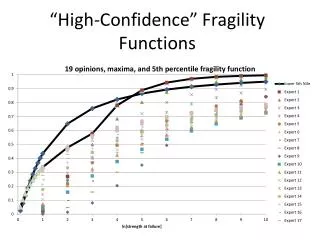
Procedure The results of the test may then be graphed, with the percent hemolysis plotted on the ordinate (vertical axis) and the sodium chloride concentration on the abscissa (horizontal axis) as shown in Figure (shows normal range).
Discussion • Presence of hemolytic organisms in the sample. • Severe anemia or other conditions with fewer RBCs available for testing . • Recent blood transfusion. • Old sample. • The pH of the blood-saline mixture is important and should be 7.4. • If anticoagulated blood is used for test, use only heparin as the anticoagulant, in order to avoid adding more salts to the blood such as Oxalate, EDTA, or citrate.
Discussion • Completely fill the collection tube and invert it gently several times to mix the sample and anticoagulant thoroughly. • Handle the sample gently to prevent accidental hemolysis.