Download
1 / 10
100 likes | 177 Views
Special Sensory Reception. Equilibrium (balance) and Hearing. Middle ear. Internal ear (labyrinth). External ear. Auricle (pinna). Helix. Lobule. External acoustic meatus. Tympanic membrane. Pharyngotympanic (auditory) tube. Anatomy of the Ear. Three parts of the ear:

E N D
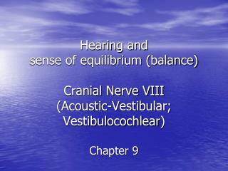
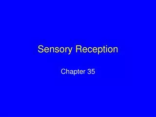
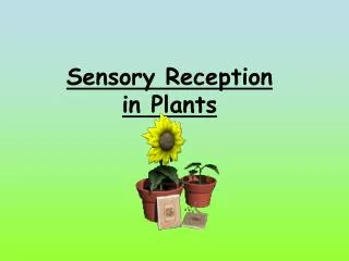
Special Sensory Reception Equilibrium (balance) and Hearing
Middle ear Internal ear (labyrinth) External ear Auricle (pinna) Helix Lobule External acoustic meatus Tympanic membrane Pharyngotympanic (auditory) tube
Anatomy of the Ear • Three parts of the ear: • External (outer) ear • Includes the auricle (pinna) which surroundes the entrance to the external acoustic meatus (ear canal) • The auricle protects the ear canal and collects and funnels sound into the ear canal • Ceruminous glands are along the external acoustic meatus and secrete a waxy material that helps prevent the entry of foreign objects and insects • The external acoustic meatus ends at the tympanic membrane (eardrum) which separates the inner and middle ear
Anatomy of the Ear • Middle ear (tympanic cavity) • Air-filled chamber • Communicates with the superior portion of the pharynx (nasopharynx). This connection is called the auditory tube (pharyngotympanic tube or Eustachian tube) • Enables equilization of pressure on either side of the eardrum • Contains the auditory ossicles (3 tiny bones together) • Connect the tympamun with the inner ear • The 3 bones are the malleus, incus, stapes • Act as levers that conduct the vibrations to the inner ear
Oval window (deep to stapes) Semicircular canals Entrance to mastoid antrum in the epitympanic recess Malleus (hammer) Vestibule Incus (anvil) Auditory ossicles Vestibular nerve Stapes (stirrup) Cochlear nerve Tympanic membrane Cochlea Round window Pharyngotympanic (auditory) tube
Anatomy of the Ear • Internal (inner) ear • Provides the senses of hearing and equilibrium • Protected by a bony labyrinth • Surrounds and protects the membranous labyrinth • Subdivided into 3 parts: • Vestibule – contains sacs that provide sensations of gravity and linear acceleration • Semicircular canals – receptors here are stimulated by rotation of the head • Cochlea – receptors here provide the sense of hearing • The receptors of the inner ear are the hair cells • Communicate with sensory neurons by continually releasing small quantities of neurotransmitters
Superior vestibular ganglion Inferior vestibular ganglion Temporal bone Semicircular ducts in semicircular canals Facial nerve Vestibular nerve Anterior Posterior Lateral Cochlear nerve Cristae ampullares in the membranous ampullae Maculae Spiral organ (of Corti) Utricle in vestibule Cochlear duct in cochlea Saccule in vestibule Stapes in oval window Round window
Equilibrium • 2 aspects of equilibrium: • Dynamic equilibrium • Aids us in maintaining our balance when the head and body are moved suddenly • Static equilibrium • Maintains our posture and stability when the body is motionless • All equilibrium sensations are provided by hair cells
Hearing • Receptors of the cochlear duct provide us with the sense of hearing • The receptor responsible are hair cells similar to those in the vestibule and semicircular canals • Sound energy is converted in air to pressure pulses which stimulate hair cells along the cochlear spiral • The frequency (pitch) of the percieved sound is determined by which part of the cochlear duct is stimulated • The intensity (volume) is determined by how many hair cells at that location are stimulated
Steps in Hearing • Sound waves arrive at the tympanic membrane • Movement of the tympanic membrane causes displacement of the auditory ossicles • The movement of the stapes at the oval window establishes pressure waves in the preilymph of the vestibular duct • The pressure waves distort the basilar membrane on their way to the round window of the tympanic duct • Vibration of the basilar membrane causes vibration of hair cells against the tectorial membrane • Information about the region and intensity of stimulation is relayed to the CNS over the cochlear branch of the cranial nerve VIII