Download
1 / 33
340 likes | 530 Views
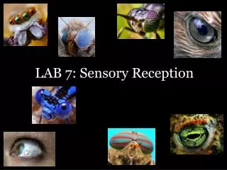
Sensory Reception. Chapter 35. Sensory Systems. The means by which organisms receive signals from the external world and internal environment Many animals can sense stimuli that humans cannot. Sensory Receptors. Convert the energy of a stimulus into action potentials. Mechanoreceptors

E N D
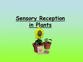
Sensory Reception Chapter 35
Sensory Systems • The means by which organisms receive signals from the external world and internal environment • Many animals can sense stimuli that humans cannot
Sensory Receptors Convert the energy of a stimulus into action potentials Mechanoreceptors Thermoreceptors Pain receptors Chemoreceptors Osmoreceptors Photoreceptors
Assessing a Stimulus • Action potentials don’t vary in amplitude • Brain tells nature of stimulus by: • Particular pathway that carries the signal • Frequency of action potentials along an axon • Number of axons recruited
Recordings of Action Potentials Figure 35.3 Page 609
Sensory Adaptation A decrease in response to a stimulus being maintained at constant strength
Somatic Sensations • Touch • Pressure • Temperature • Pain • Motion • Position
Somatosensory Cortex Figure 35.4 Page 610
Receptors in Skin • Free nerve ending • Ruffini ending • Pacinian corpuscle • Bulb of Krause • Meissner’s corpuscle Figure 35.5 Page 611
Referred Pain • Sensations of pain from internal organs may be wrongly projected to part of the skin surface • Heart attack can be felt as pain in skin above the heart and along the left shoulder and arm
Taste • A special sense • Chemoreceptors • Five primary sensations: • sweet, sour, salty, bitter, and umami Figure 35.8 Page 612
Smell • A special sense • Olfactory receptors • Receptor axons lead to olfactory lobe olfactory bulb receptor cell Figure 35.7 Page 612
Balance and Equilibrium • In humans, organs of equilibrium are located in the inner ear • Vestibular apparatus semicircular canals utricle saccule vestibular apparatus Figure 35.9bPage 613
Acceleration-Deceleration • Moving in response to gravity, otoliths bend projections of hair cells and stimulate the endings of sensory neurons otoliths hair cell membrane vestibular nerve Figure 35.9bPage 613
Dynamic Equilibrium • Rotating head movements cause pressure waves that bend a gelatinous cupula and stimulate hair cells inside it cupula Figure 35.9cPage 613
Properties of Sound • Ear detects pressure waves • Amplitude of waves corresponds to perceived loudness • Frequency of waves (number per second) corresponds to perceived pitch
Anatomy of Human Ear stirrup anvil auditory nerve hammer auditory canal eardrum cochlea Fig. 35.11a Page 614
Sound Reception • Sound waves make the eardrum vibrate • Vibrations are transmitted to the bones of the middle ear • The stirrup transmits force to the oval window of the fluid-filled cochlea
Sound Reception • Movement of oval window causes waves in the fluid inside cochlear ducts oval window (behind stirrup) scala vestibuli Figure 35.11bPage 615 eardrum round window scala tympani
Sound Reception hair cells in organ of Corti lumen of cochlear duct tectorial membrane basilar membrane to auditory nerve lumen of scala tympani Figure 35.11cPage 615
Vision • Sensitivity to light does not equal vision • Vision requires two components • Eyes • Capacity for image formation in the brain
Invertebrate Eyes Limpet ocellus ommatidium cuticle epidermis lens Compound eye of a deerfly sensory neuron Figures 35.13 & 35.14Pages 616 & 617 Land snail eye
Human Eye sclera retina choroid iris fovea optic disk lens pupil cornea part of optic nerve aqueous humor ciliary muscle Figure 35.17Page 618 vitreous body
Pattern of Stimulation • Light rays pass through lens and converge on retina at back of eye • The image that forms on the retina is upside down and reversed right to left compared with the stimulus • Brain accounts for this during processing
Pattern of Stimulation Figure 35.18Page 619
Visual Accommodation • Adjustments of the lens • Ciliary muscle encircles lens • When this muscle relaxes, lens flattens, moves focal point farther back • When it contracts, lens bulges, moves focal point toward front of eye
Organization of Retina • Photoreceptors lie at the back of the retina, in front of a pigmented epithelium • For light to reach the photoreceptors, it must pass layers of neurons involved in visual processing
Organization of Retina • Signals from photoreceptors are passed to bipolar sensory neurons, then to ganglion cells Figure 35.21aPage 620
The Photoreceptors • Rods • Contain the pigment rhodopsin • Detect very dim light, changes in light intensity • Cones • Three kinds; detect red, blue, or green • Provide color sense and daytime vision
Receptive Fields signals to oscilloscope • Restricted areas that influence the activity of individual sensory neurons • Response of neuron to orientation of bar time (sec) Figure 35.22Page 621
Retina to Brain lateral geniculate nucleus visual cortex optic nerve retina Figure 35.31Page 621
Disorders of the Eye (1) • Color blindness • Focusing problems • Nearsightedness and farsightedness • Eye diseases • Trachoma • Histoplasmosis • Herpes simplex infection
Disorders of the Eye (2) • Age-related problems • Cataracts • Macular degeneration • Glaucoma • Injuries • Retinal detachment