Download
1 / 63
670 likes | 1.05k Views
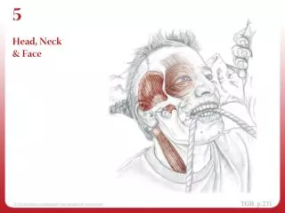
Injuries to: The Head, Neck and Face. Anatomy Review. Skull The skull has 8 cranial bones & 14 facial bones. Cranial bones have articulations called suture joints. The bones of the skull form a rigid housing for the brain

E N D
Anatomy Review Skull • The skull has 8 cranial bones & 14 facial bones. • Cranial bones have articulations called suture joints.
The bones of the skull form a rigid housing for the brain • These bones are held together by special articulations known as SUTURE JOINTS • The joints are not fully ossified until the age of 20-30 • This anatomic structure still provides a protective outer structure for the brain.
Anatomy Review (cont.)The Scalp • Soft tissue structures protect the cranium and the brain.
5 layers: • Skin • Dense connective tissue • Galea Aponeurotica (broad, flat tendon) • Loose connective tissue • Periosteum
Meninges The Meninges • Located underneath cranial bones, consisting the dura mater, arachnoid, and pia mater.
Meninges (cont.) • Dura mater is dense and highly vascularized. • Strong, Protective • Arachnoid (middle layer) is less dense and avascular. • Sub-arachnoid space contains cerebrospinal fluid (CSF). • CSF cushions the brain and spinal cord from external forces. • Pia mater (innermost layer) is thin, delicate, and highly vascularized. • More susceptible to trauma
Central Nervous System Central Nervous System (CNS) • Brain (encephalon) and spinal cord compose the CNS. • CNS is protected by meninges, cranium, and vertebrae. • CNS consists of gray and white matter and weighs 3 to 3.5 lbs (adult). • Brain has three basic components – cerebrum, cerebellum, and brain stem. • Neural impulses travel to and from the CNS via 12 pairs of cranial nerves and 31 pairs of spinal nerves.
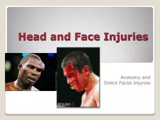
The Face The Face • The face is composed of skin placed over underlying bones. - Subcutaneous muscles, cartilage, and fat provide minor protection. • Several areas of the face are prone to injury, particularly orbits of the eyes, nasal bones, and mandible.
In Your Notebooks: • How many Cervical vertebrae are there?
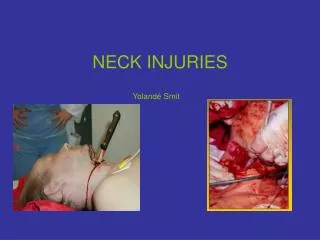
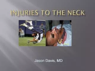
The Neck The Neck (cervical spine) • The 7 cervical vertebrae provide support for the head and protection for the spinal cord.
The first cervical vertebra (C-1) is called the atlas. The atlas articulates with the occipital bone to form R and L atlanto-occipital joints. The second cervical vertebra (C-2) is called the axis. The skull and C-1 articulate as a unit with C-2 to form the atlantoaxial joint. The Neck (cont.)
What is “Spearing”? • Why is Spearing dangerous? • What type of force is produced by Spearing? • Explain what position the neck is put in when spearing and how this relates to injury.
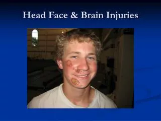
Head Injuries in Sports Even minor head trauma can result in serious injury. • Brain tissue is unable to repair itself. • Any tissue loss results in some level of permanent disability. • Severe injuries can result in death. • Coaches can learn to recognize head injuries and render first aid when necessary.
Mechanisms of Head Injury Direct mechanism of injury involves a blow to the head that causes injury at impact site (coup injury) or on the opposite side of the skull from impact (contracoup injury). Indirect injury to the head results from damaging forces traveling from other parts of the body. Treat every head injury as if there is a neck injury and vice versa.
Concussions A concussion is “a clinical syndrome characterized by immediate and transient impairment of neurologic function secondary to mechanical forces.” • Symptoms include unconsciousness, disorientation, headache, amnesia (anterograde or retrograde), dizziness, and disequilibrium.
Concussions (cont.) Cantu classification • Grade 1 (mild) involves no amnesia but are difficult to identify. • Grade 2 (moderate) involves loss of consciousness for less than 1 minute and/or PTA lasting longer than 30 minutes. • Grade 3 (severe) involves loss of consciousness for more than a minute and PTA lasting more than 24 hours.
Second Impact Syndrome (SIS) can be a serious problem. Results when an athlete with a head injury receives another head injury before the symptoms of the initial injury have resolved. Involves rapid, catastrophic brain swelling. SIS can result in death. Any athlete sustaining a head injury, no matter how minor, should be referred to a physician before being cleared to return to participation. Second Impact Syndrome
Head Injuries in Sports Cranial injury: • Involves the bones of the skull. • May also have associated soft tissue injury. • More severe forms of cranial injury involve depressed skull fractures - Involves bone fragments being pushed into the cranial region.
Head Injuries in Sports (cont.) Intracranial Injury: • These injuries are potentially life threatening. • Majority result from blunt trauma to the head. • Disruption of blood vessels results in intra-cranial bleeding (hematoma) and swelling within the cranium.
Intracranial Injuries Major forms of intracranial injury include: • Epidural hematoma. • Subdural hematoma. • Intracerebral hematoma. • Cerebral contusion. Epidural hematoma develops quickly due to arterial bleeding, while subdural hematoma develops slowly due to venous bleeding. Some degree of permanent neurologic damage and even death can result.
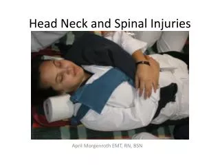
Initial Check Always assume a neck injury has also occurred. • Check vitals first. • Note body and limb positions, as well as helmet, face mask, and mouth guard positions. • If unconscious, attempt to arouse and note approx. time of injury. Immobilize head and neck immediately; do not remove athlete’s helmet.
Initial Check (cont.) • If unconscious, detect breathing by: • Listening near the athlete’s face for typical breathing sounds. • Looking for movements of the thorax and/or abdomen. If no signs of circulation are present, begin CPR and summon EMS.
Physical Examination The physical exam must include assessments of: • Consciousness or unconsciousness. • Extremity strength (if conscious) without moving the neck. • Mental function (if conscious). • Eye signs and movements. • Neck pain. • Neck musculature spasm.
Physical Exam (cont.) If head injury is suspected: • Do NOT remove helmet. • Do NOT move the athlete. • Do NOT use ammonia capsules to revive athlete. • Do NOT rush through physical exam.
Physical Exam (cont.) If athlete is conscious: • Perform grip strength tests. • Check bilateral strength by asking athlete to dorsiflex feet. • Check for sensations on both sides of body by pinch tests. • Monitor athlete’s eyes by checking pupil sizes and response to light, and eyes’ ability to follow moving object side to side. • Note loss of peripheral vision or jerking eyeball movements. • Palpate neck for deformity, moving from base of skull to bottom of neck.
Physical Exam (cont.) Based on these observations, determine level of consciousness. • Athlete with grade 1 concussion should be able to walk with assistance. • Athlete with grade 2 or 3 concussion should not be moved. • Monitor vital signs. • Summon EMS. Any athlete with a concussion should be removed from field of play and examined by a physician.
Quick Neurological Tests Romberg’s Test Finger-to-Nose Test Courtesy of Ron Pfeiffer
Cervical Spine Injuries • Cervical injuries can occur in almost any sport. • Majority occur in football, rugby, ice hockey, soccer, diving, and gymnastics. • Catastrophic injuries are rare – less than 2 in 100,000 of all neck injuries reported in the United States.
Cervical Spine Injuries (cont.) • Mechanisms of injury include hyperflexion, hyperextension, rotation, lateral flexion, and axial loading. • Muscle strains in the neck rarely involve neurologic damage. • Brachial plexus injuries can produce significant but transient symptoms. • Serious injuries occur when intact vertebra or fragments of fractured vertebra are displaced or an intervertebral disk ruptures and places pressure on spinal cord or nerve roots.
Mechanism of Cervical Spine Injuries • Any forced movement of cervical spine can result in injury. • Most cervical spine injuries result from an axial load. • Spearing in football produces axial load (NCAA prohibited technique in 1976). • In 2005, there was an increased emphasis on proper technique to eliminate spearing and minimize neck injuries.
General Treatment Guidelines Coaching personnel must take great care when conducting an examination of an athlete suspected of having a neck injury. • Neck sprains • Neck strains • Neck fractures and dislocations
Initial Treatment of Neck Injury • Medical care team leader supervises the process. • Determine if the athlete is conscious. If unconscious, check airway, breathing, and pulse (circulation). • Stabilize the head and neck. • Summon EMS. • Continue monitoring “ABCs.”
If conscious, question the athlete regarding extremity numbness or loss of feeling, weakness, and/or neck pain. If athlete reports the inability to move a limb or limbs or significant strength loss, stabilize head and neck and summon EMS. Initial Treatment of Injury Guidelines
Initial Treatment of Neck Injury (continued) If EMS arrival is delayed, place the injured athlete on a properly constructed spine board. This requires the coordinated effort of at least 5 people.
Removal of Athlete’s Helmet • Management of the helmeted player is a major issue. • Football head and face protective equipment create special problems. • In cases involving a neck injury, a football helmet provides means of cervical immobilization. Coaches should not remove the helmet.
Football Face Mask Removal • If airway must be established, removal of the face mask is necessary. • Cut the clips with a device like the “Trainer’s Angel.” • Once the clips are removed, the face mask can be rolled up, and out of the way of the airway. • If Trainer’s Angel is not available, removal of screws that hold the clips with a screwdriver is an option. Courtesy of Ron Pfeiffer
Injuries to the Maxillofacial Region • Maxillofacial injuries include those to the jaw, teeth, eyes, ears, nose, throat, facial bones, and skin. • Modern protective equipment has reduced significantly the incidence of these injuries. Such equipment includes: • Mouth guards. • Protective eye wear. • Face shields.
Dental Injuries • The human jaw has 32 teeth. • Teeth are vulnerable to external blows that are common in many sports. • Teeth are secured by cementum and periosteum.
Dental Injuries (cont.) • Majority of dental injuries result from direct blows that result in tooth displacement or avulsion, a tooth fracture, or fracture of jaw or other facial bones. • When rendering first aid, take precautions to avoid bloodborne pathogens. • When examining dental injuries: • Can athlete open and close mouth w/o pain? • What is the general symmetry of the teeth? • Are there any irregularities in adjacent teeth? • Is there bleeding, especially along gum line?
Dental Injuries (cont.) Loosened tooth: • Gently push back into place. • If tooth is avulsed, place in sterile saline and refer athlete to dentist or physician immediately. High-risk sports should require use of mouth guard. • Required for high school football players since 1966; NCAA followed suit in 1974. • Three types of mouth guards are stock, mouth-formed, and custom fitted.
Eye Injuries • Eye consists of a ball-like structure housed within the orbit. • Globe is filled with vitreous body. • The posterior interior surface is covered by the retina. • Most of the eyeball is encased in the sclera.
Eye Injuries (cont.) Approximately 40,000 sports-related eye injuries occur annually in the United States. Majority of eye injuries are preventable. Eye injuries in the U.S. are on the increase; basketball, baseball, and softball are leading sports for eye injuries. Racket sports are also responsible for eye injuries. Proper position of the fingers for an initial examination of the eye
Eye Injuries (cont.) Two categories of eye injuries are contusional and penetrating. • Contusional injuries vary in severity from simple corneal abrasions to major injuries such as rupture of the eye, fracture of orbit, or combinations of the two. Detached retina can also occur. • Penetrating injuries are less common than contusional injuries. Protective eyewear is strongly recommended.
Eye Injuries (cont.) Initial Check and Treatment Guidelines • Majority of injuries are simple corneal abrasions or small foreign object in eye. • Hold upper eyelid away from anterior eye. • Small foreign bodies usually found below lower eyelid or in the medial canthus. • Visible foreign object can be removed with a moist cotton swab; if imbedded, cover both eyes and transport to medical facility.
Eye Injuries (cont.) • If nothing can be seen in the eye, injury is probably a corneal abrasion. • Contusions may result in hemorrhage around the eye known as a “black eye.” • More severe cases may involve bleeding into the anterior eye (“hyphema”) and orbital blowout. Refer to medical facility immediately. • Symptoms of orbital blowout include eye pain, double vision (diplopia), and obvious bleeding within the eye.