Download
1 / 37
410 likes | 703 Views
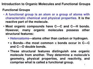
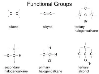
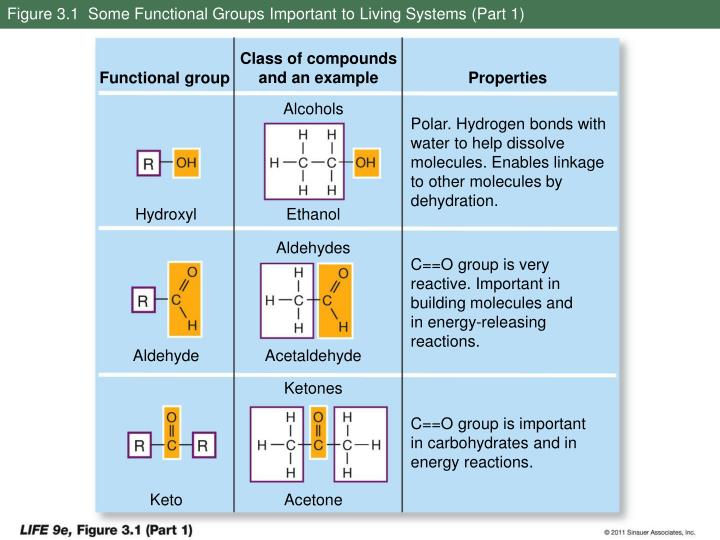
Figure 3.1 Some Functional Groups Important to Living Systems (Part 1). Class of compounds and an example. Functional group. Properties. Alcohols. Polar. Hydrogen bonds with water to help dissolve molecules. Enables linkage to other molecules by dehydration. Hydroxyl. Ethanol. Aldehydes.

E N D
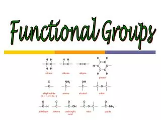
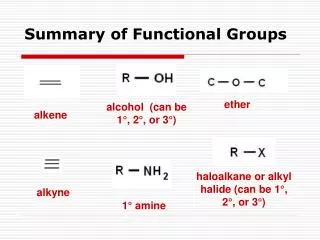
Figure 3.1 Some Functional Groups Important to Living Systems (Part 1) Class of compounds and an example Functional group Properties Alcohols Polar. Hydrogen bonds with water to help dissolve molecules. Enables linkage to other molecules by dehydration. Hydroxyl Ethanol Aldehydes C==O group is very reactive. Important in building molecules and in energy-releasing reactions. Aldehyde Acetaldehyde Ketones C==O group is important in carbohydrates and in energy reactions. Keto Acetone
Figure 3.1 Some Functional Groups Important to Living Systems (Part 2) Class of compounds and an example Functional group Properties Carboxylic acids Acidic. Ionizes in living tissues to form —COO– and H+. Enters into dehydration synthesis by giving up —OH. Some carboxylic acids important in energy-releasing reactions. Carboxyl Acetic acid Amines Basic. Accepts H+ in living tissues to form —NH3 Enters into dehydration synthesis by giving up H+. Amino Methylamine
Figure 3.1 Some Functional Groups Important to Living Systems (Part 3) Class of compounds and an example Functional group Properties Organic phosphates Negatively charged. Enters into dehydration synthesis by giving up —OH. When bonded to another phosphate, hydrolysis releases much energy. Phosphate 3-Phosphoglycerate Thiols By giving up H, two —SH groups can react to form a disulfide bridge, thus stabilizing protein structure. Sulfhydryl Mercaptoethanol
Butane Isobutane
Figure 3.2 Optical Isomers Molecule Mirror image Mirror image Hand Asymmetrical carbon atoms
Figure 3.3 Substances Found in Living Tissues Proteins (polypeptides) Macromolecules Nucleic acids Water Carbohydrates (polysaccharides) Ions and small molecules Lipids
Figure 3.4 Condensation and Hydrolysis of Polymers Condensation Hydrolysis Monomer
Side chain α carbon Amino group Carboxyl group
Figure 3.5 A Disulfide Bridge Cysteine molecules in polypeptide chain Side chains
Figure 3.6 Formation of Peptide Linkages Amino group Carboxyl group Peptide linkage N terminus (+H3N) C terminus (COO–)
Figure 3.7 The Four Levels of Protein Structure (Part 1) Peptide linkage Amino acid monomers Primary structure
Figure 3.7 The Four Levels of Protein Structure (Part 2) Hydrogen bond Secondary structure β pleated sheet α helix Hydrogen bond
Figure 3.7 The Four Levels of Protein Structure (Part 3) Tertiary structure Quaternary structure β pleated sheet Subunit 1 Subunit 2 Hydrogen bond Disulfide bridge α helix Subunit 3 Subunit 4
Figure 3.8 Three Representations of Lysozyme Space-filling model Stick model Ribbon model β pleated sheet β pleated sheet α helix α helix
Figure 3.9 Primary Structure Specifies Tertiary Structure (Part 1) Chemically denature functional ribonuclease, disrupting disulfide bridges and other intramolecular interactions that maintain the protein’s shape, so that only primary structure (i.e., the amino acid sequence) remains. Once denaturation is complete, remove the disruptive chemicals. α helix β pleated sheet Disulfide bridge Denatured protein
Figure 3.9 Primary Structure Specifies Tertiary Structure (Part 2) When the disruptive agents are removed, three-dimensional structure is restored and the protein once again is functional.
Figure 3.10 Quaternary Structure of a Protein α subunits β subunits Heme
Figure 3.11 Noncovalent Interactions Between Proteins and Other Molecules Molecule 1 Molecule 2
Figure 3.12 Chaperones Protect Proteins from Inappropriate Binding Denatured protein “Lid” HSP60 “cage”
Figure 3.13 From One Form of Glucose to the Other Aldehyde group Hydroxyl group α-D-glucose β-D-glucose Straight-chain form Intermediate form
Figure 3.14 Monosaccharides Are Simple Sugars Three-carbon sugar Five-carbon sugars (pentoses) Glyceraldehyde Ribose Deoxyribose Six-carbon sugars (hexoses) α-mannose α-galactose Fructose
Figure 3.15 Disaccharides Form by Glycosidic Linkages (Part 1) α-1,2 glycosidic linkage Formation of α linkage α-D-glucose Fructose α-D-glucose Fructose Sucrose
Figure 3.15 Disaccharides Form by Glycosidic Linkages (Part 2) α-1,4 glycosidic linkage Formation of α linkage α-D-glucose α-D-glucose β-D-glucose β-D-glucose Maltose
Figure 3.15 Disaccharides Form by Glycosidic Linkages (Part 3) β-1,4 glycosidic linkage Formation of α linkage β-D-glucose β-D-glucose β-D-glucose β-D-glucose Cellobiose
Figure 3.16 Representative Polysaccharides (Part 1) Molecular structure Cellulose Starch and glycogen
Figure 3.16 Representative Polysaccharides (Part 2) Macromolecular structure Linear (cellulose) Branched (starch) Highly branched (glycogen) Polysaccharides in cells
Figure 3.17 Chemically Modified Carbohydrates (Part 1) Sugar phosphate Phosphate groups Fructose Fructose 1,6 bisphosphate
Figure 3.17 Chemically Modified Carbohydrates (Part 2) Amino sugars Glucosamine Galactosamine
Figure 3.17 Chemically Modified Carbohydrates (Part 3) N-acetyl group Chitin Glucosamine N-acetylglucosamine Chitin
Figure 3.18 Synthesis of a Triglyceride Glycerol (an alcohol) Ester linkage + 3 Fatty acid molecules Triglyceride
Figure 3.19 Saturated and Unsaturated Fatty Acids (Part 1) Palmitic acid Oxygen Hydrogen Carbon
Figure 3.19 Saturated and Unsaturated Fatty Acids (Part 2) Linoleic acid
Figure 3.20 Phospholipids (Part 1) Phosphatidylcholine Positive charge Choline Hydrophilic head Negative charge Phosphate Glycerol Hydrophobic tail Hydrocarbon chains
Figure 3.20 Phospholipids (Part 2) Phospholipid bilayer Water Hydrophilic “heads” Hydrophobic fatty acid “tails” Hydrophilic “heads” Water
Figure 3.21 -Carotene is the Source of Vitamin A Central double bond -carotene Vitamin A Vitamin A
Figure 3.22 All Steroids Have the Same Ring Structure Cholesterol Vitamin D2 Cortisol Testosterone
Fatty acid Alcohol Ester linkage