Download
1 / 34
340 likes | 343 Views
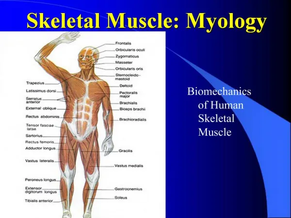
Myology. Muscles of the Anterior Neck. Muscles of the Neck Overview. Muscle of neck are divided into two groups: Anterior Superficial (2) Hyoids Infrahyoids (4) Suprahyoids (4) Scalenes (3) Deep (4) Posterior Superficial (4) Deep (4)

E N D
Myology Muscles of the Anterior Neck
Muscles of the Neck Overview • Muscle of neck are divided into two groups: • Anterior • Superficial (2) • Hyoids • Infrahyoids (4) • Suprahyoids (4) • Scalenes (3) • Deep (4) • Posterior • Superficial (4) • Deep (4) • Note: Some sources divide neck into anterior, posterior, & lateral.
Muscles of Neck Overview • Functionality • Since these muscles cross the joints of the cervical spine, they can move the neck at the cervical spinal joints • If a muscle also crosses the atlanto-occipital joint (C0/C1) then it can move the head upon the neck.
Muscles of Neck Overview • General Rules: • If a muscle crosses the neck posteriorly, it can extend the neck at the cervical spinal joints. • If a muscle crosses the neck anteriorly, it can flex the neck at the cervical spinal joints. • If a muscle crosses the neck laterally, it can laterally flex the neck at the cervical spinal joints. • If a muscle wraps around the neck, it can cause rotation of the neck at the cervical spinal joints.
Muscles of the Anterior Neck – Superficial (2) • Platysma : • By function it is primarily a muscle of facial expression i.e. innervated by CN VII. • Platysma of one side blends with contralateral side and other facial muscles in lower face. • Sternocleidomastoid (SCM): • Since it attaches to sternum, SCM is considered an accessory muscle of respiration.
O: Subcutaneous Fascia of Superior Chest I: Mandible and subcutaneous fascia of lower face A: Draws up the skin of superior chest and neck, creating ridges in neck skin. Assists in drawing the lip laterally and depresses the mandible N: CN VII (Facial nerve) Platysma Palpation: Page 138
Sternocleidomastoid (SCM) O: Sternal Head: manubrium Clavicular Head: Medial clavicle I: Mastoid process Actions: Bilateral contraction: flexion of the neck. Unilateral contraction results in Lateral flexion of neck/head and Contralateral rotation of neck/head N: Spinal accessory nerve (CN XI) Palpation: Page 141
Muscles of the Anterior Neck – Infrahyoids (4) • All 4 infrahyoid muscles are located below the hyoid bone i.e. the pull hyoid bone inferiorly when contracted. • All hyoid muscle are important in moving and/or fixating the hyoid bone. These functions are necessary for chewing, swallowing, & speech. • Sternohyoid: • “Sterno” refers to sternum • “hyoid” refers to hyoid bone • Sternothyroid: • “thyroid” refers to thyroid cartilage • Thyrohyoid • Omohyoid: • “Omo” refers to the shoulder
Sternohyoid O: Posterior aspect of the manubrium and medial clavicle I: Inferior Hyoid A: Depression of hyoid N: Ansa cervicalis of the cervical plexus Palpation: page 147
Sternothyroid O: Posterior Sternum and 1st costal cartilage I: Thyroid Cartilage A: Depression of thyroid cartilage N: Ansa cervicalis of the cervical plexus Palpation: page 150
Thyrohyoid O: Thyroid Cartilage I: Hyoid (inferior aspect) A: Depression of hyoid and Elevation of thyroid cartilage N: CN XII (Hypoglossal nerve) Palpation: page 152
Omohyoid O: Inferior Belly: Superior angle of the scapula Superior Belly: Clavicle via the central bound to the clavicle I: Inferior belly: Clavicle (via the central bound to the clavicle) Superior belly: hyoid A: Depression of hyoid N: Ansa cervicalis of the cervical plexus Palpation: page 155
Muscles of the Anterior Neck – Suprahyoids (4) • Digastric: • “Di” means two; “gastric” means belly • External carotid lies inferior and deep to anterior belly • Stylohyoid: • External carotid lies inferior and deep to stylohyoid • Mylohyoid: • “mill” refers to molar teeth • Geniohyoid: • “genio” refers to chin
Digastric O: Posterior belly: mastoid notch of temporal bone Anterior belly: Inner surface of the mandible I: Hyoid (via the central tendon) A: Elevation of hyoid, depression of the mandible, and retraction of the mandible. N: anterior belly: CN V (Trigeminal nerve) posterior belly CN VII (Facial nerve) Palpation: page 158
Stylohyoid O: Styloid process of temporal bone I: Hyoid Actions: Elevation of hyoid N: CN VII (Facial nerve) Palpation: page 161
Mylohyoid O: Entire inner surface of mandible (this muscle forms the muscular floor of the mouth) I: Hyoid A: Elevation of hyoid and depresses the mandible N: CN V (Trigeminal nerve) Palpation: page 164
Geniohyoid O: Inner surface of mandible, deep to the mylohyoid I: Hyoid A: Elevation of hyoid N: CN XII (Hypoglossal nerve) Palpation: page 167
Muscles of the Anterior Neck – Scalenes (3) • As a group, they attach superiorly from cervical TP's to inferiorly on the 1st and 2nd ribs • As a group, scalenes flex and laterally flex the neck • Nerve supply , C3- C8 • By reverse muscles action, the scalenes can elevated the 1st & 2nd rib i.e. they are also considered accessory muscle of respiration. • A fourth muscle, the scalenus minimus (Sibson's muscle), is sometimes present behind the lower portion of the scalenus anterior. • They originate from the transverse processes of the cervical vertebrae of C2 to C7 and insert onto the first and second ribs. Thus they are called the lateral vertebral muscles.
The scalene muscles have an important relationship to other structures in the neck. • The brachial plexus and subclavian artery pass between the anterior and middle scalenes. • The subclavian vein and phrenic nerve pass anteriorly to the anterior scalene as the muscle crosses over the first rib. • The phrenic nerve is oriented vertically as it passes in front of the anterior scalene, while the subclavian vein is oriented horizontally as it passes in front of the anterior scalene muscle.
Clinical relevance • Since the nerves of the brachial plexus pass through the space between the anterior and middle scalene muscles, that area is sometimes targeted with the administration of regional anesthesia by physicians. • The nerve block, called an interscalene block, may be performed prior to arm or shoulder surgery. They also act as accessory muscles of respiration when additional effort is required.
Anterior Scalene O: Anterior tubercles of the TP’s of C3 – C6 I: 1st Rib A: Bilateral contraction: flexion of the neck. Unilateral contraction causes lateral flexion and contralateral rotation of the neck. Reversed muscle action causes Elevation of 1st rib N: Ventral rami of the cervical spinal nerves Palpation: page 173
Middle Scalene O: Posterior tubercles of the TP’s of C2 to C7 I: 1st Rib A: Bilateral contraction: flexion of the neck. Unilateral contraction causes lateral flexion of the neck. Reversed muscle action causes Elevation of 1st rib N: Ventral rami of the cervical spinal nerves 3 and 4 Palpation: page 176
Posterior Scalene O: Posterior tubercles of the TP’s of cervical spine I: 2nd Rib A: Unilateral contraction causes lateral flexion of the neck. Reversed muscle action causes Elevation of 2nd rib N: Ventral rami of the cervical spinal nerves 3 and 4. Palpation: page 179
Scalenus minimus • The Scalenusminimus is an anterior muscle of the neck. • Anatomical Attachments: According to Travell and Simons, this muscle is absent in a large percentage of the population. It lies posterior to the subclavian artery, underneath the inferior aspect of the Scalenus anterior. • Origin: Extends from the transverse process of the 7th cervical vertebrae • Insertion: Attaches to the fascia supporting the dome of the pleura and inner border of the 1st rib. • Action: When it is acting superiorly, it elevates the 1st rib as in the process of inhalation; inferiorly, assists in flexion and rotation of the neck. • Nerve Supply: Cervical nerve 7. • Vascular supply: Muscular branches of the ascending Cervical artery.
The Scalene Group • Scalenes, as well as SCM, are often injured during MVA called whiplash. • Also known as cervical acceleration deceleration (CAD) injury
Muscles of the Anterior Neck – Deep Prevertebral Group (4) • Called prevertebral muscles since they lie directly on the cervical spine vertebral bodies. • Important at fixating (stabilizing) and neck/head while talking, swallowing, coughing, & sneezing LongusColli: • Has 3 parts: superior oblique, inferior oblique, & vertical • Considered to be a strong neck flexor • LongusCapitis • Rectus Capitis Anterior • Rectus CapitisLateralis
Longus Colli O: Bodies of the C3-T3 vertebrae I: TP’s and Bodies of the C1-C6 vertebrae A: Bilateral contraction causes weak flexion of neck. Unilateral contraction causes lateral flexion and contralateral rotation of the neck. N: Ventral rami of the cervical spinal nerves For the purpose of HS 113, this muscle is not palpable
Longus Capitis O: TP’s of C3 – C5 I: Basilar portion of the occiput A: Bilateral contraction causes Flexion of head/neck. Unilateral contraction causes Lateral flexion of head/neck N: Ventral rami of the cervical spinal nerves For the purpose of HS 113, this muscle is not palpable
Rectus Capitis Anterior O: TP of the Atlas (C1) I: Inferior surface of the basilar portion of the occiput A: Flexion of head N: Ventral rami of the cervical spinal nerves For the purpose of HS 113, this muscle is not palpable
Rectus Capitis Lateralis O: TP of the Atlas (C1) I: Inferior surface of the Occiput A: Lateral flexion of head N: Ventral rami of the cervical spinal nerves For the purpose of HS 113, this muscle is not palpable