Download
1 / 80
820 likes | 1.04k Views
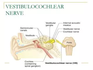
Sensory-motor transformations in vestibular processing Day 1: Linear Systems Kathleen E. Cullen and Maurice Chacron, Dept of Physiology, McGill University. The Vestibular System. The vestibular system is phylogenetically the oldest part of the inner ear:

E N D
Sensory-motor transformations in vestibular processing Day 1: Linear Systems Kathleen E. Cullen and Maurice Chacron, Dept of Physiology, McGill University
The Vestibular System The vestibular system is phylogenetically the oldest part of the inner ear: It is situated in the petrous part of the temporal bone, and is not only in close proximity to the cochlea but is continuous with the scala media.
Function of the Vestibular System Semicircular canals - sense angular rotation Otoliths - sense linear acceleration • Provide information about head motion relative to space and gravity to: • 1) Stabilize the visual axis (VOR) • 2) Maintain head and body posture (VCR and vestibulospinal reflexes) • Compute spatial orientation or ‘sense of balance’ • 4) Navigation
Function of the Vestibular System i. The VOR, ii. Posture and balance, and iii. Higher order vestibular processing
Central Vestibular Processing for the VOR Central Pathways: Vestibular Nuclei Slow phase direction = ____________ Quick phase direction = ___________ So, head velocity direction was _____________ Slow phase direction = left Quick phase direction = right So, head velocity direction = right
Sensorimotor transformations: VOR 1. Overview of Eye Movements - VOR 2. Motor Control of Eye Movements : Mechanical Constraints 3. The Vestibular System 3.1) Signal Processing by Vestibular Sensors i. Mechanical Analysis of the Semicircular Canals ii. Hair Cells and Afferent responses 3.2) Central Vestibular Processing for the VOR i. Central Pathways (Vestibular Nuclei) ii. Neuronal Pathway: Model of the VOR
How does the brain generate appropriate motor commands to move the eyes to align the axis of gaze with an object of interest?
How does the brain generate appropriate motor commands to move the eyes to align the axis of gaze with an object of interest? What are mechanical properties of the eye and surrounding tissues? Why Study Eye Movements? - No joints in system - Constant inertia (negligible)
Mechanics of Eye Movements What are the mechanics of the Oculomotor Plant? Plant: devise which produces the final output For eye movements = 1) eye muscles, 2) orbital tissues, 3) globe What is the output? Eye Movement What is the input? Muscle tension
Control System Analysis • A system is represented as: • Where x(t) is the input, and y(t) is the output. These are signals that very as a function of time. • The goal of an engineer, is to design S, so that x results in y. • The Neurobiologist already has S, and controls x, observes y • Then tries to guess what S is.
The VOR as an Example System Where: H(t) is head velocity: here a step of velocity and Ė (t) is slow-phase eye velocity (note H(t)and Ė (t) are short hand for dH/dt, dE/dt) So in this case the problem, is to find S, for the VOR
Mechanical System Analysis • For example, to understand how you move your eye, • First, consider some examples of mechanics to relate force to eye movement: • Apply a force F to a spring of stiffness K, stretch it to length L. • Hooks Law says: F = kL
Mechanical System Analysis 2) Apply a force (F) to a system characterized by a pure viscosity (of coefficient r). A good example is a hypodermic syringe. If you push at a constant force, the plunger moves at a constant velocity dL/dt, such that: F = r dL/dt
Mechanical System Analysis 3) Put these 2 elements in series (this is a simplified muscle model): This is called a visco-elasticity. The force is shared by the elasticity (kL) and the viscosity (r dL/dt) so: F = kL + r dL/dt This is a first order differential equation and if our “system” was a visco-elasticity, solving this equation for a given input should produce the observed output.
Mechanical System Analysis: 4) Now add a mass to the system: From Newton’s law of motion F = m d2L/dt2, whered2L/dt2 is acceleration The system is now described by: F = kL + r dL/dt + m d2L/dt2 (i.e. a second order differential equation)
Mechanics of Eye Movements How does mechanical analysis in the previous slides relate to the Oculomotor Plant? Plant: devise which produces the final output For eye movements = 1) eye muscles, 2) orbital tissues, 3) globe What is the output? Eye Movement What is the input? Muscle tension, but hard to measure. We can measure motoneuron drive to muscles
How does the brain generate appropriate motor commands to move the eyes to align the axis of gaze with an object of interest? Step 1: Record Eye movements Suction contact lens used to apply forces and loads to the eye to understand the Mechanical properties of the eye and surrounding tissue. FR(t) = b + kE(t − td) + rE˙(t − td) + uË(t − td) −cFR’ David Robinson 1964
How does the brain generate appropriate motor commands to move the eyes to align the axis of gaze with an object of interest? Step 2: Record Motor and Premotor Neurons Dave Sparks 2002
Standard Classification of 5 Types of Eye Movements Classically eye movements grouped into 5 types Extraocularmotoneurons participate in all types of eye movements and their response dynamics can (largely) be predicted by the mechanics of the eye.
The Brainstem motor visual motor A given extraocular motoneuron participates in all classes of eye movements .
How does the brain generate appropriate motor commands to move the eyes to align the axis of gaze with an object of interest? Van Gisbergen et al. 1981 Sylvestre and Cullen 1999 FR(t) = b + kE(t − td) + rE˙(t − td) + uË(t − td) −cFR’
Description of MN discharge rate Recall: 1)Fr = Ro + KE + rĖ ↓ ↓ 2)Fr (s) = KE(s) + r s E(s) H(s) = E(s)/Fr(s) = (1/K)/ [(r/K)s +1] The time constant: e = r/k
Description of MN discharge rate 3)Fr – Ro = KE + rĖ if e = r/k E(t) = R(1- e –t/e) E(t) = R(1- e –1) if t = e = R(1 – 1/e) = R(1 –1/2.7) = R x .63 If : r =1, K =5, then e = 250 ms.. Consider a saccadic eye movement, the eye Dynamics with a step command of FR would be too slow. Saccades can be on target in less than 100ms.
Analysis of Motoneuron Signals Pulse Need an extra “burst” (pulse) in MN command signal in order to complete saccade in a shorter time (i.e. overcome viscous drag). Step Also need tonic activity after saccade (step) in order hold eye at new position (i.e. overcome elastic restoring forces). Note, The pulse resembles velocity + The step resembles position
Neural Circuit: Controlling Saccades Tonic activity after saccade (step) generated by the oculomotor neural integrator
Neural Circuit: Controlling Saccades Tonic activity after saccade (step) generated by the oculomotor neural integrator Current model of the neural integrator based on experimental findings in Species ranging from monkeys to Zebrafish. Miri et al, Nature NS., 2011
Linearity and Superposition: For example, the MN equation added the results from 3 experiments, together. Assumption: The system is linear. If we put in 2 signals, the output is the same as as the sum of the response of each alone.
The VOR as an Example System Where: H(t) is head velocity: here a step of velocity and Ė (t) is slow-phase eye velocity (note H(t)and Ė (t) are short hand for dH/dt, dE/dt) So in this case the problem, is to find S, for the VOR
Sensorimotor transformations: VOR So, far we have considered 1. Overview of Eye Movements - VOR 2. Motor Control of Eye Movements : Mechanical Constraints 3. The Vestibular System 3.1) Signal Processing by Vestibular Sensors i. Mechanical Analysis of the Semicircular Canals ii. Hair Cells and Afferent responses 3.2) Central Vestibular Processing i. Central Pathways (Vestibular Nuclei) ii. Neuronal Pathway: Model of the VOR
Standard Classification of 5 Types of Eye Movements Classically eye movements grouped into 5 types Extraocularmotoneurons participate in all types of eye movements and their response dynamics can (largely) be predicted by the mechanics of the eye.
Function of the Vestibular System Semicircular canals - sense angular rotation Otoliths - sense linear acceleration • Provide information about head motion relative to space and gravity to: • 1) Stabilize the visual axis (VOR) • 2) Maintain head and body posture (VCR and vestibulospinal reflexes) • Compute spatial orientation or ‘sense of balance’ • 4) Navigation
Organization of the Vestibular System Anatomy: there are 2 types of sensors on each side of the head. 1) Otoliths (linear acceleration) → saccule Macula → utricle 2) Semicircular canals (angular acceleration) → horizontal → superior Ampula → posterior (crista) Note: Entire system is continuous with scala media of the cochlea via the ductus reunions.
Function of the Vestibular System i. The VOR, ii. Posture and balance, and iii. Higher order vestibular processing
Function of the VOR Gaze Stabilization via the Vestibulo-ocular Reflex More effective than vision since response latency is very short! Huterer and Cullen, J. Neurophys., 2002
Function of the VOR Gaze Stabilization via the Vestibulo-ocular Reflex VOR Gain OKN Gain Accordingly, VOR is more effective than visually driven OKN at higher frequenceis
Mechanical Analysis of the Semicircular Canals - The 3 canals are ~ at right angles to each other. - Each of the 3 planes lie approximately in the pulling direction of one of the pairs of extraocular muscles Horizontal → horizontal for normal resting posture. Superior Posterior subtend 45o relative to the sagittal and frontal plane. Each canal consists of • 1) A circular fluid path • 2) Ampulla → crista – hair cells • → cupula – elastic membrane (water tight)
Mechanical Analysis of the Semicircular Canals Receptor Cells All hair cells are oriented in the same direction for each canal. Mechanism: Head rotates → fluid is left behind → ampulla pushes against it → bends cilia.
Mechanical Analysis of the Semicircular Canals The cupula is deflected by the movement of the endolymph, which occurs during head motion. The following sequence of events occurs: 1) the head turns 2) the endolymph tends to remain stationary due to inertial forces 3) therefore the endolymph moves relative to the canal (in the opposite direction of head motion).
Mechanical Analysis of the Semicircular Canals Stimulus = Angular acceleration But Over the frequency range of normal head movements (i.e. > .01Hz). The very ↑viscous small → properties of diameter the fluid (0.3mm) This is mathematically equivalent to ∫ (integration) ↓ Thus, the system functions as an angular speedometer (hair cell output →rotational speed) CNS → 3 canals = speed of head in 3D
Hydrodynamic analysis of the canals predicted that the relationship between the angular displacement of the endolymph (ε(t)) and the head’s angular acceleration ((t)) is: d ε2/dt2 + d ε(t)/dt + ε(t) = (t) Where: is the effective moment of inertia of the endolymph. is a damping constant that reflects the viscous drag exerted by the canal wall as the endolymph flows past it, and Is a elastic restoring factor related • The dynamics of this equation are governed by two time constants, • a long one (τ1= / = 5s) and • a short one (τ2= / =.004s).
Hydrodynamic analysis of the canals predicted that the relationship between the angular displacement of the endolymph (ε(t)) and the head’s angular acceleration ((t)) is: d ε2/dt2 + d ε(t)/dt + ε(t) = (t) Where: is the effective moment of inertia of the endolymph. is a damping constant that reflects the viscous drag exerted by the canal wall as the endolymph flows past it, and Is a elastic restoring factor related H(s) = Ε(s)/(s) H(s) = 1/ [( / )s+1] [( / ) s+1] • The dynamics of this equation are governed by two time constants, • a long one (τ1= / = 5s) and • a short one (τ2= / =.004s).
This equation says that the movement of the endolymph in the canals is opposed by two frictional forces 1) one which arises from the viscosity of the endolymph and 2) a second which is due to the elasticity of the cupula. These opposing forces cause the movement of the endolymph (relative to the cupula) to lag head acceleration (as would be the case if the only the inertia of the endolymph were significant). Thus the receptor cells which deflect the movement of the cupula are primarily sensitive to head velocity (rather than acceleration) during most natural head movements (i.e. frequency = 0.05-20Hz). • This is shown in the next slide……………
Mechanical Analysis of the Semicircular Canals Frequency Response System is characterized in terms of gain and phase (Bode Plots). Note, in this graph a phase of 0 deg is in phase with velocity, and -90 and +90 deg are in phase with acceleration and position.
Sensorimotor transformations: VOR So, far we have considered 1. Overview of Eye Movements - VOR 2. Motor Control of Eye Movements : Mechanical Constraints 3. The Vestibular System 3.1) Signal Processing by Vestibular Sensors i. Mechanical Analysis of the Semicircular Canals ii. Hair Cells and Afferent responses 3.2) Central Vestibular Processing for the VOR i. Central Pathways (Vestibular Nuclei) ii. Neuronal Pathway: Model of the VOR
Hair Cells and Afferent Responses Two types of Hair cells Type I Hair Cells Characterized by calyx like endings of the sensory fibers. Type II Hair Cells Characterized by more conventional (bulbous) cell fiber synapses.
Hair Cells and Afferent Responses Exhibit a constant resting discharge when not stimulated 1) Bending cilia towards kinocilia excites hair cell: ↑ action potential, VIII nerve. 2) Bending cilia away from kinocilia inhibits hair cell: ↓ action potential, VIII nerve. Thus, the resting discharge (spontaneous discharge) allows the CNS to sense stimulation in 2 directions (opposite via the changein activity).
Mechanism of Mechano-Neural Transduction: similar to auditory system Role of Efferent system: not yet understood
Hair Cells and Afferent Responses • Regular Versus Irregular Afferents • Afferent innervation patterns • type II haircells - regular afferents • type I haircells - irregular affernts • Regulars: • - More regular action potentials spacing • - Lower Afferent gain and phase • - Lower Efferent response magnitude • - Lower Galvanic sensitivity
Vestibular afferent Dynamics: Afferents show a response gain increase with frequency 0.5 Hz 15 Hz Regular Irregular Sadeghi, Minor, and Cullen; J Neurophys, 2007