Download
1 / 114
1.14k likes | 1.34k Views
BIF 38: Nervous & Sensory Systems. Overview: Sensing and Acting. The star-nosed mole can catch insect prey in near total darkness in as little as 120 milliseconds It uses the 11 appendages protruding from its nose to locate and capture prey

E N D
Overview: Sensing and Acting • The star-nosed mole can catch insect prey in near total darkness in as little as 120 milliseconds • It uses the 11 appendages protruding from its nose to locate and capture prey • Sensory processes convey information about an animal’s environment to its brain, and muscles and skeletons carry out movements as instructed by the brain
Command and Control Center • The human brain contains about 100 billion neurons, organized into circuits more complex than the most powerful supercomputers • A recent advance in brain exploration involves a method for expressing combinations of colored proteins in brain cells, a technique called “brainbow” • This may allow researchers to develop detailed maps of information transfer between regions of the brain
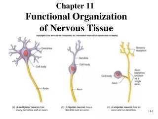
Concept 49.1: Nervous systems consist of circuits of neurons and supporting cells Each single-celled organism can respond to stimuli in its environment Animals are multicellular and most groups respond to stimuli using systems of neurons
The simplest animals with nervous systems, the cnidarians, have neurons arranged in nerve nets • A nerve net is a series of interconnected nerve cells • More complex animals have nerves • Nerves are bundles that consist of the axons of multiple nerve cells • Sea stars have a nerve net in each arm connected by radial nerves to a central nerve ring
Planarian(flatworm) (b) Sea star(echinoderm) (c) (h) Salamander(vertebrate) Figure 49.2 Eyespot Brain Brain Radialnerve Nervecords Ventralnerve cord Nervering Transversenerve Nerve net Segmentalganglia (a) Hydra (cnidarian) (d) Leech (annelid) Brain Ganglia Brain Anteriornerve ring Spinalcord(dorsalnervecord) Ventralnerve cord Brain Sensoryganglia Ganglia Longitudinalnerve cords Segmentalganglia (e) Insect (arthropod) (f) Chiton (mollusc) (g) Squid (mollusc)
Bilaterally symmetrical animals exhibit cephalization, the clustering of sensory organs at the front end of the body • Relatively simple cephalized animals, such as flatworms, have a central nervous system (CNS) • The CNS consists of a brain and longitudinal nerve cords Eyespot Brain Nervecords Transversenerve Planarian (flatworm)
Annelids and arthropods have segmentally arranged clusters of neurons called ganglia Brain Brain Ventralnerve cord Ventralnerve cord Brain Segmentalganglia Ventralnerve cord Segmentalganglia Segmentalganglia Leech (annelid) Insect (arthropod)
Nervous system organization usually correlates with lifestyle • Sessile molluscs (for example, clams and chitons) have simple systems, whereas more complex molluscs (for example, octopuses and squids) have more sophisticated systems
Ganglia Brain Anteriornerve ring Ganglia Longitudinalnerve cords Squid (complex mollusc) Chiton (“sessile” mollusc)
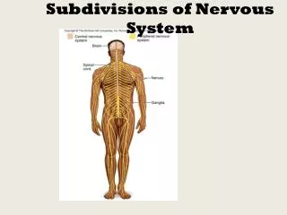
Brain • In vertebrates • The CNS is composed of the brain and spinal cord • The peripheral nervous system (PNS) is composed of nerves and ganglia Spinalcord(dorsalnervecord) Sensoryganglia Salamander (vertebrate)
Organization of the Vertebrate Nervous System • The spinal cord conveys information from and to the brain • The spinal cord also produces reflexes independently of the brain • A reflex is the body’s automatic response to a stimulus • For example, a doctor uses a mallet to trigger a knee-jerk reflex
Figure 49.3 Cell body ofsensory neuron indorsal rootganglion Gray matter Quadricepsmuscle White matter Hamstringmuscle Spinal cord(cross section) Sensory neuron Motor neuron Interneuron
Invertebrates usually have a ventral nerve cord while vertebrates have a dorsal spinal cord • The spinal cord and brain develop from the embryonic nerve cord • The nerve cord gives rise to the central canal and ventricles of the brain
Central nervoussystem (CNS) Peripheral nervoussystem (PNS) Figure 49.4 Brain Cranial nerves Spinal cord Ganglia outsideCNS Spinal nerves
The central canalof the spinal cord and the ventriclesof the brain are hollow and filled with cerebrospinal fluid • The cerebrospinal fluid is filtered from blood and functions to cushion the brain and spinal cord as well as to provide nutrients and remove wastes
The brain and spinal cord contain • Gray matter, whichconsists of neuron cell bodies, dendrites, and unmyelinated axons • White matter, whichconsists of bundles of myelinated axons Gray matter Whitematter Ventricles
Glia • Glia have numerous functions to nourish, support, and regulate neurons • Embryonic radial glia form tracks along which newly formed neurons migrate • Astrocytes induce cells lining capillaries in the CNS to form tight junctions, resulting in a blood-brain barrier and restricting the entry of most substances into the brain
CNS PNS Figure 49.6 Neuron VENTRICLE Astrocyte Cilia Oligodendrocyte Schwann cell Microglial cell Capillary Ependymal cell 50 m LM
The Peripheral Nervous System • The PNS transmits information to and from the CNS and regulates movement and the internal environment • In the PNS, afferentneurons transmit information to the CNS and efferentneurons transmit information away from the CNS
The PNS has two efferent components: the motor system and the autonomic nervous system • The motor system carries signals to skeletal muscles and is voluntary • The autonomic nervous system regulates smooth and cardiac muscles and is generally involuntary
Figure 49.7 Central NervousSystem(information processing) Peripheral NervousSystem Efferent neurons Afferent neurons Autonomicnervous system Motorsystem Sensoryreceptors Control ofskeletal muscle Sympatheticdivision Parasympatheticdivision Internaland externalstimuli Entericdivision Control of smooth muscles,cardiac muscles, glands
The autonomic nervous system has sympathetic, parasympathetic, and enteric divisions • The sympathetic division regulates arousal and energy generation (“fight-or-flight” response) • The parasympatheticdivision has antagonistic effects on target organs and promotes calming and a return to “rest and digest” functions • The enteric division controls activity of the digestive tract, pancreas, and gallbladder
Figure 49.8 Sympathetic division Parasympathetic division Action on target organs: Action on target organs: Constricts pupilof eye Dilates pupil of eye Inhibits salivarygland secretion Stimulates salivarygland secretion Sympatheticganglia Constrictsbronchi in lungs Relaxes bronchiin lungs Cervical Slows heart Accelerates heart Stimulates activityof stomach andintestines Inhibits activity ofstomach and intestines Thoracic Inhibits activityof pancreas Stimulates activityof pancreas Stimulates glucoserelease from liver;inhibits gallbladder Stimulatesgallbladder Lumbar Stimulatesadrenal medulla Promotes emptyingof bladder Inhibits emptyingof bladder Sacral Promotes erectionof genitalia Promotes ejaculationand vaginal contractions Synapse
The vertebrate brain is regionally specialized Specific brain structures are particularly specialized for diverse functions These structures arise during embryonic development
Figure 49.10 Eye Input from nervesof ears Reticular formation Input from touch,pain, and temperaturereceptors
Brain structures in child and adult Embryonic brain regions Figure 49.9b Cerebrum (includes cerebral cortex, whitematter, basal nuclei) Telencephalon Forebrain Diencephalon (thalamus, hypothalamus,epithalamus) Diencephalon Mesencephalon Midbrain (part of brainstem) Midbrain Metencephalon Pons (part of brainstem), cerebellum Hindbrain Myelencephalon Medulla oblongata (part of brainstem) Diencephalon Cerebrum Mesencephalon Metencephalon Midbrain Midbrain Diencephalon Myelencephalon Hindbrain Pons Medullaoblongata Spinal cord Forebrain Cerebellum Telencephalon Spinal cord Child Embryo at 5 weeks Embryo at 1 month
Figure 49.9c Left cerebralhemisphere Right cerebralhemisphere Cerebral cortex Corpus callosum Cerebrum Basal nuclei Cerebellum Adult brain viewed from the rear
Figure 49.9d Diencephalon Thalamus Pineal gland Brainstem Hypothalamus Midbrain Pituitary gland Pons Medullaoblongata Spinal cord
Arousal and Sleep • The brainstem and cerebrum control arousal and sleep • The core of the brainstem has a diffuse network of neurons called the reticular formation • This regulates the amount and type of information that reaches the cerebral cortex and affects alertness • The hormone melatonin is released by the pineal gland and plays a role in bird and mammal sleep cycles
Sleep is essential and may play a role in the consolidation of learning and memory • Dolphins sleep with one brain hemisphere at a time and are therefore able to swim while “asleep”
Figure 49.11 Key Low-frequency waves characteristic of sleep High-frequency waves characteristic of wakefulness Time: 1 hour Time: 0 hours Location Lefthemisphere Righthemisphere
Biological Clock Regulation • Cycles of sleep and wakefulness are examples of circadian rhythms, daily cycles of biological activity • Mammalian circadian rhythms rely on a biological clock, molecular mechanism that directs periodic gene expression • Biological clocks are typically synchronized to light and dark cycles • In mammals, circadian rhythms are coordinated by a group of neurons in the hypothalamus called the suprachiasmatic nucleus (SCN) • The SCN acts as a pacemaker, synchronizing the biological clock
Emotions • Generation and experience of emotions involve many brain structures including the amygdala, hippocampus, and parts of the thalamus • These structures are grouped as the limbic system • The limbic system also functions in motivation, olfaction, behavior, and memory • Generation and experience of emotion also require interaction between the limbic system and sensory areas of the cerebrum • The structure most important to the storage of emotion in the memory is the amygdala, a mass of nuclei near the base of the cerebrum
Figure 49.13 Thalamus Hypothalamus Olfactorybulb Hippocampus Amygdala
Figure 49.14 Amygdala Nucleus accumbens Happy music Sad music
Drug Addiction and the Brain’s Reward System • The brain’s reward system rewards motivation with pleasure • Some drugs are addictive because they increase activity of the brain’s reward system • These drugs include cocaine, amphetamine, heroin, alcohol, and tobacco • Drug addiction is characterized by compulsive consumption and an inability to control intake
Addictive drugs enhance the activity of the dopamine pathway • Drug addiction leads to long-lasting changes in the reward circuitry that cause craving for the drug
Nicotinestimulatesdopamine-releasingVTA neuron. Inhibitory neuron Figure 49.23 Opium and heroindecrease activityof inhibitoryneuron. Dopamine-releasingVTA neuron Cocaine andamphetaminesblock removalof dopaminefrom synapticcleft. Cerebralneuron ofrewardpathway Rewardsystemresponse
The cerebral cortex controls voluntary movement and cognitive functions
The cerebrum, the largest structure in the human brain, is essential for awareness, language, cognition, memory, and consciousness • Four regions, or lobes (frontal, temporal, occipital, and parietal), are landmarks for particular functions
Figure 49.15 Motor cortex(control ofskeletal muscles) Somatosensory cortex(sense of touch) Frontal lobe Parietal lobe Prefrontal cortex(decision making,planning) Sensory associationcortex (integration ofsensory information) Visual associationcortex (combiningimages and objectrecognition) Broca’s area(forming speech) Temporal lobe Occipital lobe Auditory cortex (hearing) Visual cortex(processing visualstimuli and patternrecognition) Cerebellum Wernicke’s area(comprehending language)
Language and Speech • Studies of brain activity have mapped areas responsible for language and speech • Broca’s area in the frontal lobe is active when speech is generated • Wernicke’s area in the temporal lobe is active when speech is heard • These areas belong to a larger network of regions involved in language
Figure 49.16 Max Hearingwords Seeingwords Min Speakingwords Generatingwords
Lateralization of Cortical Function • The two hemispheres make distinct contributions to brain function • The left hemisphere is more adept at language, math, logic, and processing of serial sequences • The right hemisphere is stronger at pattern recognition, nonverbal thinking, and emotional processing • The differences in hemisphere function are called lateralization • Lateralization is partly linked to handedness • The two hemispheres work together by communicating through the fibers of the corpus callosum
Information Processing • The cerebral cortex receives input from sensory organs and somatosensory receptors • Somatosensory receptors provide information about touch, pain, pressure, temperature, and the position of muscles and limbs • The thalamus directs different types of input to distinct locations
Adjacent areas process features in the sensory input and integrate information from different sensory areas • Integrated sensory information passes to the prefrontal cortex, which helps plan actions and movements • In the somatosensory cortex and motor cortex, neurons are arranged according to the part of the body that generates input or receives commands
Figure 49.17 Frontal lobe Parietal lobe Shoulder Upper arm Trunk Elbow Trunk Head Forearm Knee Neck Leg Hip Hip Wrist Elbow Hand Forearm Fingers Hand Fingers Thumb Thumb Eye Neck Nose Brow Eye Face Genitalia Lips Toes Face Teeth Gums Lips Jaw Jaw Tongue Pharynx Tongue Primarysomatosensorycortex Primarymotor cortex Abdominalorgans