Download
1 / 25
250 likes | 263 Views
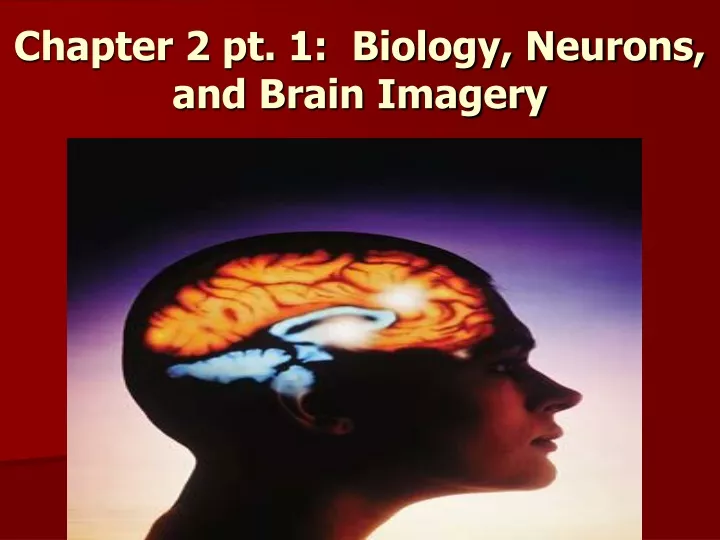
Chapter 2 pt. 1: Biology, Neurons, and Brain Imagery. Review: What Link Do Biological Psychologists Attempt To Study?. Biological Psychologists study the link between: The basic building block of the nervous system is called the neuron or a nerve cell. Parts of the Neuron.

E N D
Review: What Link Do Biological Psychologists Attempt To Study? • Biological Psychologists study the link between: • The basic building block of the nervous system is called the neuron or a nerve cell.
Parts of the Neuron • 1. Dendrites: branching extensions that receive incoming messages and conduct messages toward the cell body. • 2. Soma: is the cell body, which contains the nucleus.
Parts of A Neuron • 3. Axon: extension of a neuron which takes messages from the soma to other neurons; is the longest part of the neuron. • 4. Terminal Buttons: located on end of the axon that release neurotransmitters to communicate with other neurons. • 5. Myelin Sheath:a layer of fatty cells segmentally encasing the fibers of many axons which allows faster transmission speeds in neurons
How Does A Neuron Communicate? • Action Potential: neural impulse or brief electrical charge that travels down an axon at speeds as fast as 200 mph. Is enacted when sense receptors feel something. Is considered an “ALL OR NOTHING” response. • Resting Potential: refers to the neuron when it is not active. Is negatively charged. • Threshold: refers to the minimal level of stimulation required for a neural impulse to fire.
Neuron Communication With Other Neurons • In order for one neuron to communicate with another it must pass a junction or gap called the synapse between the axon which is sending the signal and the dendrite which is receiving the signal. • At the ends of the axon, the terminal buttons release neurotransmitters: which are chemical messengers that bind together neurons and influence whether another neural impulse will take place.
Types of Neurotransmitters…These Are Tough!! • 1. Acetylcholine: vital role in learning and memory but most well known for its presence in allowing muscle contraction. • Shortage may lead may lead to Alzheimer’s disease or muscular disorders.
Types of Neurotransmitters…These Are Tough!! • 2.Serotonin: affects mood, hunger, and arousal. • Shortage may lead to depression. • 3. Dopamine: influences movement, attention, and emotion. • Excess may lead to schizophrenia and Parkinson’s disease.
Types of Neurotransmitters…These Are Tough!! • 4. Norepinephrine: helps control alertness and arousal when you are scared or excited. • 5. Endorphins: called the “morphine within” because of its link to pain control and pleasure.
Types of Neurotransmitters…These Are Tough!! • GABA: helps relax and calm down the body. • Shortage may cause Anxiety or Epilepsy.
Agonists vs. Antagonists • Agonists are chemicals that mimic the effects of a neurotransmitter. Ex: • Antagonists are chemicals that block the transmission of a neurotransmitter. Ex: • https://www.youtube.com/watch?v=haNoq8UbSyc
The Body’s Nervous System • Nervous System: is your electrochemical communication center; consists of the 1. central nervous system and the nerves within your 2. peripheral nervous system.
2 Divisions: CNS vs. PNS • Central Nervous System (CNS): contains the brain and spinal cord. • Peripheral Nervous System (PNS): contains the sensory and motor neurons that connect the CNS to the rest of the body. Is Divided into Autonomic and Somatic.
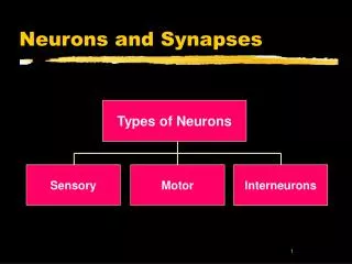
3 Types of Neurons • 1. Sensory Neurons: neurons that carry info from the sensory receptors (in the PNS) to the brain and spinal cord (in the CNS). Also called Afferent Neurons. • 2.Interneurons: neurons within the CNS that internally communicate between sensory and motor neurons. • 3. Motor Neurons: take information from the CNS to the muscles and glands within the PNS. Also called Efferent Neurons.
Nervous system Peripheral Central (brain and spinal cord) Autonomic (controls self-regulated action of internal organs and glands like The heart and lungs) Skeletal (controls voluntary movements of skeletal muscles) Sympathetic (arousing) Parasympathetic (calming) Divisions of the Nervous System
Automatic Actions=Are Simple Reflexes • Reflex: a simple, autonomic, inborn response to a sensory stimulus. Spinal cord in charge and brain NOT involved.
How to Study the Brain • Lesion: natural or experimentally damaged tissue of the brain used to study portions of the brain.
Studying the Brain • EEG (Electroencephalogram): an amplified recording of the waves of electrical activity that sweep across the brain’s surface; these waves are measured by electrodes placed on the scalp -Used in sleep studies.
Studying the Brain • CT (computed tomograph) Scan (CAT SCAN):a series of x-ray photographs taken from different angles and combined by computer into a composite representation of a slice through the body. Also called CAT scan. • Allows one to see soft-tissue structures.
Studying the Brain • PET (positron emission tomograph) Scan:a visual display of brain activity that detects where a radioactive form of glucose goes while the brain performs a given task. • Shows brain activity.
Studying the Brain • MRI: (magnetic resonance imaging):a technique that uses magnetic fields and radio waves to produce computer – generated images that distinguish among different types of soft tissue; allows us to see structures within the brain.