Download

1 / 23
240 likes | 521 Views
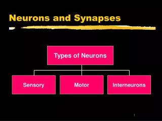
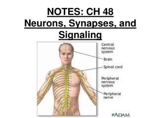
Neurons and Synapses. Types of Neurons. Sensory. Motor. Interneurons. Sensory Neurons. INPUT From sensory organs to the brain and spinal cord. Brain. Drawing shows a somatosensory neuron Vision, hearing, taste and smell nerves are cranial, not spinal. Sensory Neuron. Spinal Cord.

E N D
Neurons and Synapses Types of Neurons Sensory Motor Interneurons
Sensory Neurons • INPUT Fromsensory organs to the brain and spinal cord. Brain Drawing shows a somatosensory neuron Vision, hearing, taste and smell nerves are cranial, not spinal Sensory Neuron Spinal Cord
Brain Sensory Neuron Spinal Cord Motor Neuron Motor Neurons • OUTPUTFrom the brain and spinal cord To the muscles and glands.
Brain Sensory Neuron Spinal Cord Motor Neuron Interneurons • Interneurons carry information between other neurons only found in the brain and spinal cord.
The cell body • Round, centrally located structure • Contains DNA • Controls protein manufacturing • Directs metabolism • No role in neural signaling • Contains the cell’s Nucleus
Dendrites • Information collectors • Receive inputs from neighboring neurons • Inputs may number in thousands • If enough inputs the cell’s AXON may generate an output
Dendritic Growth • Mature neurons generally can’t divide • But new dendrites can grow • Provides room for more connections to other neurons • New connections are basis for learning
Axon • The cell’s output structure • One axon per cell, 2 distinct parts • tubelike structure branches at end that connect to dendrites of other cells
Myelin Sheath Myelin sheath • White fatty casing on axon • Acts as an electrical insulator • Not present on all cells • When present increases the speed of neural signals down the axon.
How neurons communicate • Neurons communicate by means of an electrical signal called the Action Potential • Action Potentials are based on movements of ions between the outside and inside of the cell • When an Action Potential occurs a molecular message is sent to neighboring neurons
Outside of Cell K+ Na+ Cl- Cell Membrane in resting state K+ Na+ Cl- A- Inside of Cell Ion concentrations
K+ Na+ Cl- Outside of Cell Cell Membrane at rest Na+ - 70 mv A- K+ Cl- Inside of Cell Potassium (K+) can pass through to equalize its concentration Sodium and Chlorine cannot pass through Result - inside is negative relative to outside The Cell Membrane is Semi-Permeable
Resting Potential • At rest the inside of the cell is at -70 microvolts • With inputs to dendrites inside becomes more positive • if resting potential rises above threshold an action potential starts to travel from cell body down the axon • Figure shows resting axon being approached by an AP
Depolarization ahead of AP • AP opens cell membrane to allow sodium (NA+) in • inside of cell rapidly becomes more positive than outside • this depolarization travels down the axon as leading edge of the AP
Repolarization follows • After depolarization potassium (K+) moves out restoring the inside to a negative voltage • This is called repolarization • The rapid depolarization and repolarization produce a pattern called a spike discharge
Finally, Hyperpolarization • Repolarization leads to a voltage below the resting potential, called hyperpolarization • Now neuron cannot produce a new action potential • This is the refractory period
Dendrite Axon Cell Body Neuron to Neuron • Axons branch out and end near dendrites of neighboring cells • Axon terminals are the tips of the axon’s branches • A gap separates the axon terminals from dendrites • Gap is the Synapse
Sending Neuron Axon Synapse Terminal Synapse • axon terminals contain small storage sacs called synaptic vesicles • vesicles contain neurotransmitter molecules
Neurotransmitter Release • Action Potential causes vesicle to open • Neurotransmitter released into synapse • Locks onto receptor molecule in postsynaptic membrane
Locks and Keys • Neurotransmitter molecules have specific shapes • Receptor molecules have binding sites • When NT binds to receptor, ions enter positive ions (NA+ ) depolarize the neuron negative ions (CL-) hyperpolarize
Some Drugs work on receptors • Some drugs are shaped like neurotransmitters • Antagonists : fit the receptor but poorly and block the NT • e.g. beta blockers • Agonists : fit receptor well and act like the NT • e.g. nicotine.
Summary • 3 types of neurons • The cell membrane • Ion movements • Action potentials • Synapse • Neurotransmitters • Receptors and ions • Agonists and antagonists