Download
1 / 26
270 likes | 373 Views
Paraxial mesoderm and somitogenesis. Dóra Dávid 2018.03.08. Classification of the mesoderm. Cells migrated between ecto and endoderm during gastrulation 5 types of mesoderm exist chorda mesoderm paraxial mesoderm intermedier mesoderm lateral mesoderm head mesoderm. Somitogenesis.

E N D
Paraxial mesoderm and somitogenesis Dóra Dávid 2018.03.08.
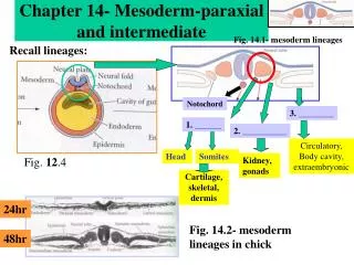
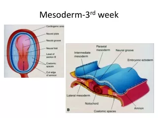
Classification of the mesoderm • Cells migrated between ecto and endoderm during gastrulation • 5 types of mesoderm exist • chorda mesoderm • paraxial mesoderm intermedier mesoderm lateral mesoderm head mesoderm
Somitogenesis somites - Somites are simmetrical, segmental structures appearing in specific timeframes lateral to the neural tube - They originate from paraxial mesoderm -During their differentiation their progenitor cell will form the: vertebrae, ribs, intervertebral discs dermis, hipodermis back, thoracic, abdominal, limb skeletal muscles
Ephrin and its receptor constitute a possible cut site for somite formation cranial • EphA4 Direction of somitogenesis caudal • Somitogenesis: repeating sequence of similar morfogenetic events • Cranio-caudal directed process
Paraxial mesoderm Humán embrióban 37-40 somita fejlődik ki Neural tube somite Paraxial mesoderm
Formation of somites • Shape: like a ball, covered by epithelial cells, inside mesenchyme • One pair of somites form during a species specific time-frame (in chick embryo it is 90 minutes) • They are identical in size • .
Clock and Wavefrontmodel • Somites form during preset time-frame, but the 7 cranial somitomer never transform into somites origin of head mesoderm, and of skeletal muscles of head • The time-adjusted cyclical process is depicted by the „Clock and Wavefront” model • Presence of oscillatorically expressing genes in paraxial mesoderm → expression interval of these genes are identical with the time frame of somite formation • FGF8 (produced by Hensen’s node and tail bud) and retinoic acid (produced by already formed somites) establish an oppository gradient in the cranio-caudal axis of the embryo • FGF8 and retinoic acid „extinguishing” each other time the expression of „oscillatory genes”
Delta-Notch are expressed at presumptive boundaries Ectoderm induction with Wnt-6 will induce the paraxis gene, thus expression changes the mesenchymal somitomer into a epithelial somite
Somites further differenciate after their formation: • Sclerotome • Syndetome • Dermomyotom
Sclerotome: Ventromedial cell group of somites. During multiple mitosis they migrate around the developing neural tube and notochord due to the effect of Shh and noggin, produced by the latters Expression of Pax-1 and Pax-9 begins secondary mesenchyme formation and production of cartilage-specific ECM molecules. Dermomyotome: Effect of ectodermal Wnt secretion differentiate it to two parts: dermatome (close to the ectoderm). Progenitors of dermotome (together with progenitors from the lateral mesoderm) together will establish the layers of dermis and hypodermis The part closer to the neural tube of the dermomyotome will be the myotome. Later it contributes to the development of skeletal muscles of the body wall, limbs and back. Cells of the dermomyotom express Pax-3 and Pax-7 Syndetome: Layer between the sclerotome and dermomyotome. Its cells will differentiate to the precursors of tendocytes
Differentiation of somites noggin
Sclerotome determination • Shh signaling from notochord/floor plate • Inducing Pax1 expression
Ventral sclerotome: corpus vertebrae, discus intervertebralis Lateral sclerotome: distal ribs Medial sclerotome (not shown): meninges and vessels (meningotome) Arthrotome: discus intervertebralis, proximal ribs Dorsal sclerotome: dorsal part of arcus vertebrae, processus spinosus Central sclerotome: ventral part of arcus vertebrae, processus transversus
Sclerotomes will split to a cranial and caudal segment further • The caudal segment is more compacted, the cranial is more loosened • Two neighbouring sclerotome’s caudal and cranial segments will form a vertebra • Motoneurons’ axons from the neural tube just can pass through the loosened cranial segment
Differentiation of sclerotome: Formation of vertebrae caudal cranial
Somites and their sclerotomes are identical but vertebrae are different... Explanation is the differential expression of HOX genes and the so called HOX-code
epithelio-mesenchymal transmission dermamyotom Dermatom ectoderm epidermis dermis dermatome hypo-dermis Neural tube neurotropin 3 (NT-3)
dermamyotome Myotom Body wall Tongue muscles Limb msucles Back muscles Progenitors of epimere will form: m. erector spinae and transversospinal muscles (epaxial muscles) Progenitors of hypomere will form: intercostal muscles, m. obliquus externus, m. obliquusinternus, m. transversus abdominis and limb muscles Noggin signal from notochord inhibits ectodermal BMP-4, instead of Pax-3 and Pax-7 myogenic factors starts to express, like MyoD or Myf5
Migration of myoblasts limb bud Migrating myoblasts BMP-4 from the lateral mesoderm extinguishes the myogenic effect, And cells start to express Pax-3 C-met: tirosine kinase receptor protein appears on the surface of myoblasts, to what HGF(hepatocyte growth factor) binds as a ligand expressed by the limb bud’s mesenchymal cells
Trunk muscles at the level of limb buds epaxial myotome Autochton back musculature no hypaxial myotome Pax-3, HGF/SF, c-met Myogenic cells for limb muscles
Between lim buds Surface ectodern dermatome epaxial myotome autochton hátizomzat sclerotome hypaxial myotome Abdominal and intercostal muscles Wolffian duct
CSIR Chicken limb bud quail chick Quail derived muscle