Download
1 / 9
90 likes | 213 Views
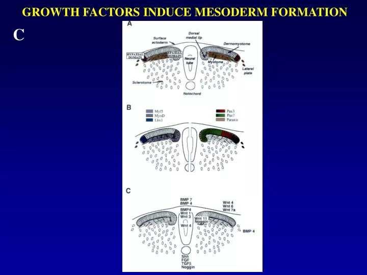
GROWTH FACTORS INDUCE MESODERM FORMATION. C. QSulf1 suppresses FGF-induced mesoderm induction in Xenopus animal cap assays.

E N D
QSulf1 suppresses FGF-induced mesoderm induction in Xenopus animal cap assays Fig. 1. Xenopus embryos were injected with QSulf1 or inactive QSulf1(C-A) mRNAs (750 pg) at the one-cell stage. Animal caps were dissected at stage 8 –9 and cultured to assess mesoderm induction and ERK1/2 activation. (A) Untreated animal cap explants form epidermis and assume a spherical shape, but caps elongate and form mesodermal tissues when induced with FGF. Expression of QSulf1, but not catalytically inactive QSulf1(C-A), blocks FGF2-induced animal cap elongation. (B) Animal caps were cultured until stage 11 before isolating RNA to assess gene expression by RT-PCR. QSulf1 blocked mesodermal gene expression (Brachyury) induced by FGF2 (30 ng_ml), whereas catalytically inactive QSulf1(C-A) had no effect. The slight reduction of Brachyury expression in this experiment was not significant when averaged in five independent experiments. EF1_ was used as a gel-loading control and whole embryo RNA was used as a positive control for expression of mesoderm genes. Wang et al., 101; 4833-4838 (2004)
QSulf1 suppresses FGF-induced mesoderm induction in Xenopus animal cap assays Fig. 1. (C and D) For assays of ERK1/2 phosphorylation, animal caps were cultured for 1 h in the presence or absence of FGF2 or FGF4 (30 ng_ml), and protein extracts were prepared for Western blot analysis using antibodies against diphosphorylated ERK1/2 (Dp-ERK1/2) and total ERK1/2 proteins. Total ERK1/2 levels were used to monitor gel loading. FGF2-induced Dp-ERK1/2 phosphorylation is suppressed by QSulf1, but not by enzymatically inactive QSulf1(C-A) (C). QSulf1 also suppressed FGF4-induced Dp-ERK1/2 activation (D). (E) Phosphorylation of ERK1/2 shown in panel C was quantified and presented as the mean with standard error from four independent experiments. The relative Dp-ERK1/2 levels were calculated by measuring the ratios of Dp-ERK1/2 to total ERK1/2, and then normalizing to an FGF2-induced level that was arbitrarily assigned a value of 10. QSulf1 repression of FGF2-induced Dp-ERK1/2 was statistically significantly different from FGF2 control (P= 0.001), whereas QSulf1(C-A) had no effect on FGF2 signaling activity (P= 0.72). Wang et al., 101; 4833-4838 (2004)
Constitutively activated FGFR1s bypass QSulf1 inhibition of FGF2 and FGF4 signaling Fig. 2. FGFR1K562E is FGFR1 with an intracellular domain mutation conferring ligand-independent constitutive activity (A and B). iFGFR1 is a membrane-docked FGFR1 receptor with a deleted extracellular domain and an added intracellular domain to promote receptor dimerization and inducible activation in response to addition of AP20187 (C). QSulf1 mRNA (2 ng) was injected alone or coinjected with FGFR1K562E mRNA (100 pg) or iFGFR1 mRNA (20 pg) into one-cell stage Xenopus embryos. Animal caps were dissected at stage 8 –9 for Western blot analysis of phoshorylated ERK1/2 (Dp-ERK1/2) and total ERK1/2 (ERK1/2) (A and C), and by RT-PCR to assay mesodermal gene expression (B). (A) Protein extracts were assayed 1 h after FGF2 treatment by Western blot analysis of Dp-ERK1/2. QSulf1 suppresses FGF2 (30 ng/ml) but not FGFR1K562E activation of Dp-ERK1/2. (B) QSulf1 suppresses FGF2, but not FGFR1K562E induction of mesodermal markers, Brachyury and MyoD. Whole-embryo samples provided a positive control for mesodermal gene expression. (C) AP20187 (1.25 M) activation of iFGFR1 receptor induces Dp-ERK1/2 activation in the presence of QSulf1. Wang et al., 101; 4833-4838 (2004)
Exogenous heparin rescues the suppression of FGF2 signaling by QSulf1 Fig. 3. QSulf1 mRNA was injected into one-cell stage Xenopus embryos (2 ng). Animal caps were dissected and cultured with soluble heparin at a concentration of 150 ng_ml. (A) Animal caps were collected after 1 h of FGF2 treatment. QSulf1 suppressed the ERK1/2 activation induced by FGF2 protein (30 ng/ml) and exogenous heparin rescued the suppression. Total ERK1/2 was assayed as a gel-loading control. (B) Animal caps were cultured until stage 11 to assess gene expression. QSulf1 suppression of mesodermal gene (Brachyury) expression was rescued by exogenous heparin. EF1was a gel-loading control and the whole embryo RNA was a positive control for meso-derm gene expression. Wang et al., 101; 4833-4838 (2004)
QSulf1-treated heparin suppresses FGF2-induced angiogenesis Fig. 4. Filters soaked with FGF2, with or without heparin, were applied to the chorioallantonic membrane of day 10 chicken embryos. Blood vessel formation on the membrane was analyzed after 3 days. (A) Application of control PBS filter did not significantly induce formation of blood vessel branches. (B) Application of FGF2 (25 ng) filter induced extensive formation of blood vessel branches. (C) Application of filter containing FGF2 and QSulf1-treated heparin (200 ng) suppressed FGF2 induction of blood vessel branches. (D) Application of filter containing FGF2 and QSulf1(C-A)-treated heparin (200 ng) did not significantly affect FGF2 induction of blood vessel branches. Arrows indicate formation of blood vessel branches. (E) Quantification of angiogenesis. An-giogenesis was scored from 1 (low) to 4 (high). Each experimental group included >14 embryos. The data are presented as the mean score and standard deviation of three independent experiments. The suppression of angiogenesis by QSulf1-treated heparin is statistically significant (P<0.05), whereas QSulf1(C-A)-treated heparin did not signifi-cantly affect FGF2-induced angiogenesis (P= 0.82). Wang et al., 101; 4833-4838 (2004)
QSulf1-treated heparin reduces the binding of FGF2 to FGFR1, but not to heparin Fig. 5. (A) QSulf1 does not affect the binding of FGF2 to heparin. Heparin beads pretreated with QSulf1 or QSulf1(C-A) mutant protein were incubated with the indicated amount of FGF2. The heparin-bound FGF2 was analyzed by Western blotting. No significant difference was detected in FGF2 binding to QSulf1- and QSulf1(C-A)-treated heparin. (B) QSulf1 treatment of heparin reduces the formation of a FGF2– heparin–FGFR1 complex. QSulf1- or QSulf1(C-A)-treated soluble heparin at the indicated concentrations was incubated with FGF2 (10 ng/ml) and FGFR1c-AP (10 ng/ml). FGFR1c-AP was immunoprecipitated with an anti-AP antibody conjugated to agarose beads, and bound FGF2 was detected by Western blotting. (C) Quantification of FGF2 bound to FGFR1c-AP in the presence of QSulf1- or QSulf1(C-A)-treated heparin. The binding of FGF2 to FGFR1c-AP in the presence of QSulf1-treated heparin was reduced 2- to 5-fold compared with the binding in the presence of QSulf1(C-A)-treated heparin. Wang et al., 101; 4833-4838 (2004)
A model of FGF signaling regulation by Sulf1 through modulating FGF ligand–receptor interaction Fig. 6. A 2:2:2 model (two ligands, two HS chains, and two receptors) was proposed by others to illustrate ternary complex formation of FGF2–HS–FGFR1 during signaling. (A) In the absence of Sulf1, sulfated cell surface HS promotes FGF ligand–receptor interaction, receptor dimerization, and activation of intracellular signaling. (B) Selective 6-O-desulfation of endogenous HS by QSulf1 reduces FGF ligand–receptor binding but has little effect on FGF ligand binding to HS. The reduced ligand–receptor interaction suppresses receptor dimerization and signaling. (C) Exogenous sulfated heparin rescues signaling by replacing QSulf1-desulfated endoge-nous cell surface HS and promoting FGF ligand–receptor interaction. (D) Sulf1 6-O-desulfated exogenous heparin has reduced affinity for FGFR1 but competitively binds to FGF2 and interferes with endogenous HS-mediated FGF2 angiogenic activity. The ‘‘P’’ in the intracellular domain of FGFR1 indicates phosphorylation and activation. Wang et al., 101; 4833-4838 (2004)
Model for epaxial and hypaxial muscle lineage determination showing that two different signaling pathways control the determination of the epaxial and hypaxial lineages in somites Figure 7 Hypaxial muscle cell progenitors are determined in the ventral lateral compartment of somites (light gray) and epaxial muscle cell progenitors are determined in the dorso-medial compartment of somites (dark gray). Our present work demonstrates the essential role of Sonic hedgehog in the determination of the epaxial muscle lineage during embryogenesis, and its function in restricting expansion of hypaxial gene expression into the ventral medial compartment. Epaxial muscle lineage determination occurs via a Myf5-dependent pathway, although a Myf5-independent route may also occur in absence of Myf5. Surface ectoderm signals, possibly Wnts (Tajbakhsh et al., 1998), are likely to control the determination of the hypaxial muscle lineage through a Myf5-dependent, but also a Myf5-independent, pathway.