Download
1 / 16
160 likes | 305 Views
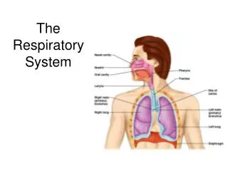
Respiratory System: - nasal cavity - Primary Bronchi, bronchioles & alveoli - Cells in alveoli. Organization of the respiratory system. Functions, movement of air (breathing); exchange oxygen for carbon dioxide Conducting portion and respiratory portion

E N D
Respiratory System: - nasal cavity - Primary Bronchi, bronchioles & alveoli - Cells in alveoli
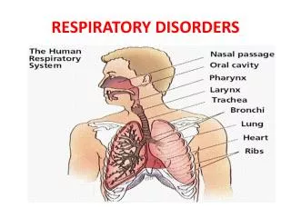
Organization of the respiratory system • Functions, movement of air (breathing); exchange oxygen for carbon dioxide • Conducting portion and respiratory portion • Conducting: epithelium, cartilage, smooth muscle, bones • Nasal and oral cavities, nasopharynx, pharynx, larynx, tracea, primary bonchi, secondary bronchi, brondhiles, terminal bronchioles.
Conducting portion • warm air, filter out bacteria, prevent dehydration, immune against infection • Olfactory epithelium
Nasal Cavity • Respiratory epithelium and keratinizing stratifed squamous epithelium • Pseudostratified ciliated epithelium also in the trachea and bronchioles (respiratory ep) • Ciliated columnar cells – secretory IgA transcytosis • Goblet cells • Basal cells • Brush cells with tall microvilli • DNES cells (diffuse neuroendocrine system)
Olfactory epithelium • OSNs • SCs • BC • Bowman’s glands • Glomeruli (1000 – 1500) around 10,000 odors
Trachea • Mucosa – respiratory epithelium • Lamina propria, loose fibroelastic tissue, lymph elements, elastic lamina • Submucosa – dense fibro-elastic, mucous and seromucous glands, lymphoid elements, BV • Adventitia – C rings, fibroelastic CT, anchors to various structures
Primary Bronchi - histological structure same as trachea
Intrapulmonary bronchi - adventitia has irregular plates of cartilage - elastic fibers - between lamina propria and submucosa - smooth muscle - form opposing spirals around bronchi - lamina propria with seromucous glands
Bronchioles - ciliated simple columnar, few goblet cells --> simple cuboidal, no goblet cells, yes Clara cells Clara cells - produce surfactant - contain cytochrome P450 enzymes - proliferate and can differentiate to epithelial cells Lamina Propria - no glands - surrounded by a loose network of helically oriented smooth muscle cells **no cartilage**
Terminal bronchioles - epithelium is cuboidal ciliated with Clara cells - one or two layers of smooth muscle Respiratory bronchioles - similar to terminal bronchioles - wall has alveolar outpouchings Alveolar Ducts - linear arrangements of alveoli
Alveolar sacs - cluster of alveoli Alveoli - - primary structural and functional unit of the respiratory system - allow for gas exchange between air and blood
Cells in alveoli: Type I pneumocytes - simple squamous epithelium - occluding junctions - basal lamina Type II pneumocytes - cuboidal cells - form occluding junctions with type I pneumocytes - contain lamellar bodies with pulmonary surfactant that is secreted by exocytosis Alveolar macrophages - phagocytose dust and bacteria
Continuous capillaries: - endothelial cells without fenestrae - fasciae occludentes Blood-Gas Barrier - surfactant and type I pneumocytes - fused basal laminae - endothelial cells
Digestive System - Oral Cavity Tongue - epidermis, taste cells, glands, muscles Lingual papillae: - filiform - no taste buds - fungiform - dorsal taste buds - foliate - taste buds in neonates, lateral furrows - circumvallate - look like a submerged donut - taste buds on sides
Taste buds - - taste pore - nerve fibers form synaptic junctions with type I, type II, type III cells - type IV are basal cells
Glands of the tongue - von Ebner’s glands - serous glands - ducts at the base of circumvallate and folate papillae