Download
1 / 139
1.39k likes | 1.6k Views
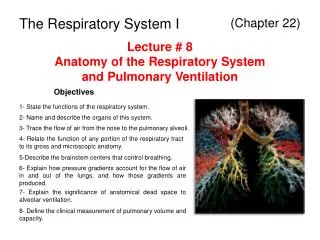
Chapter 22. Ventilation (Respiratory) System BIOL242. Overview. Respiratory anatomy Respiration (Ventilation!) Respiratory musculature Ventilation, lung volumes and capacities Gas exchange and transport O 2 CO 2 Respiratory centers Chemoreceptor reflexes Respiratory Diseases.

E N D
Chapter 22. Ventilation (Respiratory) System BIOL242
Overview • Respiratory anatomy • Respiration (Ventilation!) • Respiratory musculature • Ventilation, lung volumes and capacities • Gas exchange and transport • O2 • CO2 • Respiratory centers • Chemoreceptor reflexes • Respiratory Diseases
Oxygen • Is obtained from the air by diffusion across delicate exchange surfaces of lungs • (Which tissue lines it?) • Is carried to cells by the cardiovascular system which also returns carbon dioxide to the lungs
Functions of the Respiratory System • Supplies body with oxygen (O2)and gets rid of carbon dioxide (CO2) • Provides extensive gas exchange surface area between air and circulating blood • Moves air to and from exchange surfaces of lungs • Protects respiratory surfaces from outside environment • Produces sounds • Participates in olfactory sense
Components of the Respiratory System Figure 23–1
Organization of the Respiratory System • Upper respiratory system • Nose, nasal cavity, sinuses, and pharynx • Lower respiratory system • Larynx, trachea, bronchi and lungs
The Respiratory Tract • Conducting zone: • from nasal cavity to terminal bronchioles • conduits for air to reach the sites of gas exchange • Respiratory zone: • the respiratory bronchioles,alveolar ducts, and alveoli • sites of gas exchange
The Respiratory Epithelium Figure 23–2
Respiratory Epithelia • Changes along respiratory tract • Nose, nasal cavity, nasopharynx = pseudostratified ciliated columnar epithelium • Oropharynx, laryngopharynx = stratified squamous epitheium • Trachea, bronchi = pseudostratified ciliated columnar epithelium • Terminal bronchioles = cuboidal epithelium • Respiratory bronchioles, alveoli = simple squamous epithelium • Think about why each part has the lining that it does • For example, in alveoli • walls must be very thin (< 1 µm) • surface area must be very great (about 35 times the surface area of the body) • In lower pharynx • walls must be tough because food abrades them
TheRespiratoryMucosa • Consists of: • epithelial layer • areolar layer • Lines conducting portion of respiratory system • Lamina propria • Areolar tissue in the upper respiratory system, trachea, and bronchi (conducting zone) • Contains mucous glands that secrete onto epithelial surface • In the conducting portion of lower respiratory system, contains smooth muscle cells that encircle lumen of bronchioles
Respiratory Defense System • Series of filtration mechanisms removes particles and pathogens • Hairs in the nasal cavity • Goblet cells and mucus glands: produce mucus that bathes exposed surfaces • Cilia: sweep debris trapped in mucus toward the pharynx (mucus escalator) • Filtration in nasal cavity removes large particles • Alveolar macrophages engulf small particles that reach lungs
Upper Respiratory Tract Figure 23–3
Upper Respiratory Tract • Nose : • Air enters through nostrils or external nares into nasal vestibule • Nasal hairs in vestibule are the first particle filtration system • Nasal Cavity : • Nasal septum divides nasal cavity into left and right • Mucous secretions from paranasal sinus and tears clean and moisten the nasal cavity • Meatuses Constricted passageways in between conchae that produce air turbulence: • Warm (how?) and humidify incoming air (bypassed by mouth breathing) • trap particles • Air flow: from external nares to vestibule to internalnares throughmeatuses, then to nasopharynx
The Pharynx • A chamber shared by digestive and respiratory systems that extends from internal nares to the dual entrances to the larynx and esophagus at the C6 vertebrae • Nasopharynx • Superior portion of the pharynx (above the soft palate) contains pharyngeal tonsils; epithelium? • Oropharynx • Middle portion of the pharynx, from soft palate to epiglottis; contains palatine and lingual tonsils; communicates with oral cavity; epithelium? • Laryngopharynx • Inferior portion of the pharynx, extends from hyoid bone to entrance to larynx and esophagus
Lower Respiratory Tract • Air flow from the pharynx enters the larynx, continues into trachea, bronchial tree, bronchioles, and alveoli
Anatomy of the Larynx Figure 23–4
Cartilages of the Larynx • 3 large, unpaired cartilages form the body of the larynx (voice box) • thyroid cartilage(Adam’s apple) • hyaline cartilage • Forms anterior and lateral walls of larynx • Ligaments attach to hyoid bone, epiglottis, and other laryngeal cartilages • cricoid cartilage • hyaline cartilage • Form posterior portion of larynx • Ligaments attach to first tracheal cartilage • the epiglottis • elastic cartilage • Covers glottis during swallowing • Ligaments attach to thyroid cartilage and hyoid bone
Small Cartilages of the Larynx • 3 pairs of small hyaline cartilages: • arytenoid cartilages • corniculate cartilages • cuneiform cartilages • Corniculate and arytenoid cartilages function in opening and closing the glottis and the production of sound
Larynx Functions • To provide a patent airway • To function in voice production • To act as a switching mechanism to route air and food into the proper channels • Thyroid and cricoid cartilages support and protect the glottis and the entrance to trachea • During swallowing the larynx is elevated and the epiglottis folds back over glottis preventing entry of food and liquids into respiratory tract
Sphincter Functions of Larynx • The larynx is closed during coughing, sneezing, and Valsalva’smaneuver • Valsalva’smaneuver • Air is temporarily held in the lower respiratory tract by closing the glottis • Causes intra-abdominal pressure to rise when abdominal muscles contract • Helps to empty the rectum • Acts as a splint to stabilize the trunk when lifting heavy loads • Glottis also “closed” (covered) by epiglottis during swallowing
The Glottis Figure 23–5
Sound Production • Air passing through glottis: • vibrates vocal folds and produces sound waves • Sound is varied by: • tension on vocal folds • voluntary muscles position cartilages
Anatomy of the Trachea Figure 23–6
The Trachea • Extends from the cricoid cartilage into mediastinum where it branches into right and left bronchi • Has mucosa, submucosa which contains mucous glands, and adventitia • Adventita made up of 15–20 C-shaped tracheal cartilages (hyaline)strengthen and protect airway • Ends of each tracheal cartilage are connected by an elastic ligament and trachealis muscle where trachea contacts esophagus. Why?
The Primary Bronchi • Right and left primary bronchi are separated by an internal ridge (the carina) • Right primary bronchus • larger in diameter than the left • descends at a steeper angle
TheBronchial Tree • Formed by the primary bronchi and their branches • Each primary bronchus (R and L) branches into secondary bronchi, each supplying one lobe of the lungs (5 total) • Secondary Bronchi Branch to form tertiary bronchi • Each tertiary bronchus branches into multiple bronchioles • Bronchioles branch into terminalbronchioles: • 1 tertiary bronchus forms about 6500 terminal bronchioles
Bronchial Tree Figure 23–9
Bronchial Structure • The walls of primary, secondary, and tertiary bronchi: • contain progressively less cartilage and more smooth muscle, increasing muscular effects on airway constriction and resistance • Bronchioles: • Consist of cuboidal epithelium • Lack cartilage support and mucus-producing cells and are dominated by a complete layer of circular smooth muscle
Autonomic Control • Regulates smooth muscle: • controls diameter of bronchioles • controls airflow and resistance in lungs • Bronchodilation of bronchial airways • Caused by sympathetic ANS activation • Reduces resistance • Bronchoconstriction • Caused by parasympathetic ANS activation or • histamine release (allergic reactions)
The Bronchioles Figure 23–10
Conducting Zones Figure 22.7
Lungs Figure 23–7
The Lungs • Left and right lungs: in left and right pleural cavities • The base: • inferior portion of each lung rests on superior surface of diaphragm • Hilus • Where pulmonary nerves, blood vessels, and lymphatics enter lung • Anchored in meshwork of connective tissue
Lung Anatomy • Lungs have lobes separated by deep fissures • Right lung is wider and is displaced upward by liver. Has 3 lobes: • superior, middle, and inferior • separated by horizontal and oblique fissures • Left lung is longer is displaced leftward by the heart forming the cardiac notch. Has2 lobes: • superior and inferior • separated by an oblique fissure
Relationship between Lungs and Heart Figure 23–8
Respiratory Zone • Each terminal bronchiole branches to form several respiratory bronchioles, where gas exchange takes place (Exchange Surfaces) • Respiratory bronchioles lead to alveolar ducts, then to terminal clusters of alveolar sacs composed of alveoli • Approximately 300 million alveoli: • Account for most of the lungs’ volume • Provide tremendous surface area for gas exchange
Alveoli • Alveoliare air-filled pockets within the lungs where all gas exchange takes place • Alveolar epithelium is a very delicate, simple squamous epithelium • Contains scattered and specialized cells • Lines exchange surfaces of alveoli
Alveolar Organization Figure 23–11
Alveolar Organization • Respiratory bronchioles are connected to alveoli along alveolar ducts • Alveolar ducts end at alveolar sacs: common chambers connected to many individual alveoli • Each individual alveolus has an extensive network of capillaries and is surrounded by elastic fibers
Alveolar Epithelium • Consists of simple squamous epithelium (Type I cells) • Patrolled by alveolar macrophages, also called dust cells • Contains septal cells (Type II cells) that produce surfactant: • oily secretion containing phospholipids and proteins • coats alveolar surfaces and reduces surface tension
Alevolar problems • Respiratory Distress: difficult respiration • Can occur when septal cells do not produce enough surfactant • leads to alveolar collapse • Pneumonia: inflammation of the lung tissue • causes fluid to leak into alveoli • compromises function of respiratory membrane
Respiratory Membrane • The thin membrane of alveoli where gas exchange takes place. Consists of: • Squamous epithelial lining of alveolus • Endothelial cells lining an adjacent capillary • Fused basal laminae between alveolar and endothelial cells • Diffusion across respiratory membrane is very rapid because distance is small and gases (O2 and CO2) are lipid soluble
Blood Supply to Respiratory Surfaces • Pulmonary arteries branch into arterioles supplying alveoli with deoxy. blood • a capillary network surrounds each alveolus as part of the respiratory membrane • blood from alveolar capillaries passes through pulmonary venules and veins, then returns to left atrium with oxy. blood INTERCONNECTEDNESS
Blood Supply to the Lungs Proper • Bronchial arteries provide systemic circulation bringing oxygen and nutrients to tissues of conducting passageways of lung • Arise from aorta and enter the lungs at the hilus • Supply all lung tissue except the alveoli • Venous blood bypasses the systemic circuit and just flows into pulmonary veins • Blood Pressure in the pulmonary circuit is low (30 mm Hg) • Pulmonary vessels are easily blocked by blood clots, fat, or air bubbles, causing pulmonary embolism
Pleural Cavities and Membranes • 2 pleural cavities are separated by the mediastinum • Each pleural cavity holds a lung and is lined with a serous membrane = the pleura: • Consists of 2 layers: • parietal pleura • visceral pleura • Pleural fluid: a serous transudate thatlubricates space between 2 layers
Respiration • Refers to 4 integrated processes: • Pulmonary ventilation – moving air into and out of the lungs (provides alveolar ventilation) • External respiration – gas exchange between the lungs and the blood • Transport – transport of oxygen and carbon dioxide between the lungs and tissues • Internal respiration – gas exchange between systemic blood vessels and tissues
Gas Pressure and Volume Figure 23–13
Boyle’s Law • Defines the relationship between gas pressure and volume: P 1/V Or P1V1 = P2V2 • In a contained gas: • external pressure forces molecules closer together • movement of gas molecules exerts pressure on container
Pulmonary Ventilation Respiration: Pressure Gradients Figure 23–14