Download
1 / 44
790 likes | 2k Views
Diabetic Foot Infection. UHN AIMGP Seminar Updated Oct 2007 R. Cavalcanti. References. IDSA Guidelines: Diagnosis and Treatment of Diabetic Foot Infections. CID Oct 1, 2004; 39:885-910

E N D
Diabetic Foot Infection UHN AIMGP Seminar Updated Oct 2007 R. Cavalcanti
References • IDSA Guidelines: Diagnosis and Treatment of Diabetic Foot Infections. CID Oct 1, 2004; 39:885-910 • Singh N, Armstrong D, Lipsky B. Preventing Foot Ulcers in Patients with Diabetes. JAMA, January 12, 2005 – 293(2): 217-228 • Canadian Diabetes Association 2003 Clinical Practice Guidelines
Case • Mr. M. is a 45M who presents to AIMGP clinic for diabetes follow up. His main complain is right foot pain and swelling for one week. • What aspects of the history are important? • How about physical examination? • Which investigations would you order?
Introduction • Foot infections in patients with diabetes • Large morbidity and mortality • Frequent visits to health care professionals • #1 cause of leg amputation • Diabetic foot infections require attention to local (foot) and systemic (metabolic) issues by a multidisciplinary foot care team.
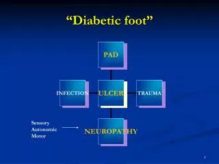
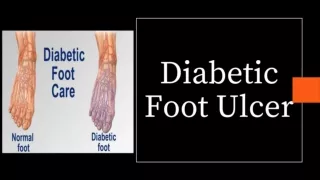
Definitions • Diabetic foot infection: any infra-malleolar infection in a person with diabetes mellitus. • Includes: paronychia, cellulitis, myositis, abscesses, necrotizing fasciitis, septic arthritis, tendinitis, and osteomyelitis.
Pathophysiology Neuropathy Trauma or Immunologic excessive pressure Disturbances lack of protective Sensation Skin breakdown Infection Peripheral Vascular Disease
Risk Factors for DM foot infection • Neuropathy • Sensory: Lack of protective sensation-> trauma • Motor: Changes in foot anatomy-> pressure points • Autonomic: Lack of sweat = dry cracked skin • Neuro-osteoarthropathy: pressure points • Peripheral vascular disease: Ischemia • Metabolic (hyperglycemia): • decreased immune defenses, poor wound healing
Patient and Health Care Delivery Factors • Patient disabilities: poor eyesight, previous amputations • Patient behaviour: compliance with preventive measures, hygiene • Health care system failures: • lack of patient education • glycemic control targets not met • poor implementation of preventive strategies
Assessment • Patient • Limb or foot • Wound
Constitutional – fever, chills, sweats, vomiting Cognitive state – confused, depressed Social situation – self neglect, non-compliance, lack of home support Neuropathy – loss of sensation Wound – pain, warmth, purulence PMHx: duration of DM, glycemic control, smoking History
Case continued • Mr. M denies any constitutional symptoms. • He has noted an ulcer over his right forefoot which is red and tender. • He has had DM1 for 30 years with retinopathy and peripheral neuropathy. • He had a left midfoot amputation 2 years ago. His glycemic control is sub-optimal. He smokes 1-2 cigarettes per day. • He is unmotivated and depressed but is worried about losing his other leg.
Physical Examination: what to look for • Vital signs – tachycardia, hypotension • Signs of volume depletion • Cognitive state – delirium, stupor, coma • Limb / foot • Biomechanics: deformities(eg charcot arthropathy) • Vascular status • Arterial – inspection: ischemia, necrosis, gangrene; palpation: pulses; special tests: ABI • Venous – edema, stasis, thrombosis • Neuropathy: light touch, vibration, monofilament pressure perception
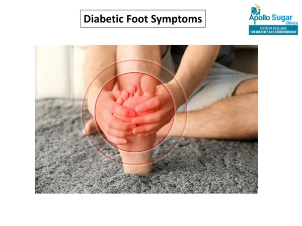
Physical Examination • Wound • Size and depth: • necrosis, gangrene, foreign body • involvement of muscle, tendon, bone, or joint – • inspect, debride, and probe the wound! • Presence, extent and cause of infection: • purulence, warmth, tenderness, induration, cellulitis, • bullae, crepitus, abscess, fasciitis, osteomyelitis
Physical Examination • Infection should be diagnosed clinically based on the presence of • Purulent secretions or • At least two of the cardinal manifestations of inflammation (redness, warmth, swelling or induration, and pain or tenderness)
Case continued … • On physical examination he is alert and oriented. Vital signs are stable. • Examination of the right foot: purulent superficial ulcer on the 5th metatarsal head with 1 cm induration and erythema around the ulcer. • There is no visible or palpable bone with probing. Peripheral pulses are palpable. • Light touch, vibration, and monofilament pressure perception is greatly diminished.
Investigations • Bloodwork – rule out hyperglycemia, DKA, hyperosmolar state • Gram staining and culture • Imaging • Radiography (2 or more) • +/- MRI to rule out osteomyelitis • Arterial Doppler +/- angiogram • ? Ultrasound to rule out deep abscesses
Investigations Note – Assess foot’s arterial supply in every patient with a diabetic foot infection (A-II). If dorsalis pedis and posterior tibialis pulses are palpable, arterial supply is generally adequate. If in doubt, other diagnostic tests are indicated (ABI, dopplers).
Principles of Therapy • Avoid prescribing antibiotics for uninfected ulcerations. • Determine the need for hospitalization (severe infection or critical limb ischemia require hospitalization) • Stabilize the patient • Restoration of fluid and electrolyte balance • Correction of hyperglycemia, hyperosmolarity, acidosis, and azotemia • Treat other exacerbating factors Note – improving glycemic control may aid in eradicating the infection and healing the wound
Principles of Therapy • Choose an antibiotic regimen • Severe infection: • start broad spectrum IV abx (ensure GPC, gram negative and anaerobic coverage) • Mild-Moderate infection: • Relatively narrow spectrum only covering aerobic GPC • No evidence for anti-anaerobic therapy • Oral therapy with highly bioavailable agents is appropriate • Mildly infected open wounds with minimal cellulitis: • Limited data support the use of topical antimicrobial therapy (B-I)
Principles of Therapy • Determine the need for surgery • Ranges from debridement to revascularization • Urgent surgical consultation for life- or limb-threatening infections (eg nec fasc, gas gangrene, compartment syndrome, critical ischemia, etc) • Formulate a wound care plan • Daily inspection • Moist wound healing environment • Removal of pressure • Debridement as needed
Principles of Therapy • Adjunctive treatment • G-CSF: does not speed healing but reduces the need for operative procedures (preliminary meta-analysis of 5 randomized trials) • Hyperbaric oxygen therapy: reserved for chronic, nonhealing ulcers. Evidence indicates it reduces the risk of major amputation related to a diabetic foot ulcer (Cochrane review)
Follow up • Careful observation of patients’ response to therapy – daily for inpatients and q2-5d for outpatients • Primary indicators of improvement: resolution of local and systemic symptoms and clinical signs of inflammation • WBC, ESR, CRP of limited use for monitoring response
Follow up • Select the definitive abx regimen: review culture and drug susceptibility results • More virulent species should always be covered, eg S. aureus, and group A or B strep) • It is not always necessary to cover less virulent species (eg CNS or enterococci) in a polymicrobial culture. If no response to empirical Rx, select agents with activity against all isolates.
Follow up • Re-evaluate the wound • Review the offloading and wound-care regimens – determine effectiveness of the regimen and patient’s compliance • Evaluate the glycemic control
Osteomyelitis • Consider OM as a potential complication of any deep or extensive ulcer, esp. if chronic or overlying a bony prominence • Suspect OM if an ulcer does not heal after at least 6 weeks of appropriate Abx therapy and off-loading • Any ulcer in which bone is visible or easily palpable with probe is likely to be OM. • Sausage toe, unexplained high WBC count or inflammatory markers are suspicious for OM
Osteomyelitis • Diagnosis: • Serial radiography (2-4 wk interval) • If typical changes: treat as OM • If c/w OM but not characteristic: • Further imaging: MRI or nuclear scan or • Continue empiric abx Rx for another 2-4 wks and repeat radiography to r/o progression of infection or • Bone biopsy • MRI is the most accurate imaging modality.
Outcome • Good clinical response to appropriate therapy in • 80-90% of mild-moderate infection • 60-80% of severe infections or cases of OM • Poor outcome associated with: • Signs of systemic infection • Inadequate limb perfusion • OM • Necrosis or gangrene • Inexperienced surgeon • Proximal location of the infection • Relapse rate: 20-30%
Prevention • Early detection of neuropathy • Educate the patient about: • Optimizing glycemic control • Using appropriate footwear at all times • Avoid foot trauma • Perform daily self-examination of the feet and report changes to health care professionals • Smoking cessation • Refer patients with severe neuropathy, substantial foot deformity, or critical ischemia
Neuropathy • Early detection of neuropathy is best accomplished in the primary care setting with a brief history and Semmes-Weinstein monofilament.
Neuropathy • History: • previous foot ulceration (RR 1.6) • prior lower extremity amputation (RR 2.8) • long duration of DM (>10 yrs, OR 3.0) • poor glycemic control (HbA1c >9%; OR 3.2) • impaired vision (acuity <20/40; RR 1.9)
Neuropathy • Screening for loss of protective sensation • Monofilament: 66-91% sen, 34-86% spe, PPV 18-39%, NPV 94-95% • Testing 8-10 anatomic sites recommended • 4 plantar sites on the forefoot identifies 90% of the patients with an insensate site • Tuning fork: less predictive of ulceration compared to monofilaments
Back to the case … • You diagnosed mild diabetic foot infection and started treatment with oral cephalexin for 2 weeks after obtaining culture. • You also counseled Mr. M. for smoking cessation and optimizing glycemic control. • You arranged a follow-up appointment in 7 days to reassess the wound and response to treatment. • Other thoughts?
Next: Algorithm for review • Please refer to these algorithm slides if needed • Otherwise just use them for reference