Download
1 / 26
410 likes | 1.09k Views
Diabetic Foot. d r. Ngr Gd Boyke A.W. Epidemiology. 40% - 60% of all non traumatic lower limb amputation 85% of diabetic related foot amputation are preceded by foot ulcer 4 out of 5 ulcer in diabetics are precipitated by trauma 4% -10% is the prevalence of foot ulcer in diabetics.

E N D
Diabetic Foot dr. NgrGdBoyke A.W.
Epidemiology • 40% - 60% of all non traumatic lower limb amputation • 85% of diabetic related foot amputation are preceded by foot ulcer • 4 out of 5 ulcer in diabetics are precipitated by trauma • 4% -10% is the prevalence of foot ulcer in diabetics
Systemic burden due to high blood sugars • damaging white cells to reduceleucocyte function. • disrupting blood vessel endothelium giving diabetic microangiopathy thereby reducing tissue perfusion • giving microorganisms an ideal environment to proliferate and infect ulcers
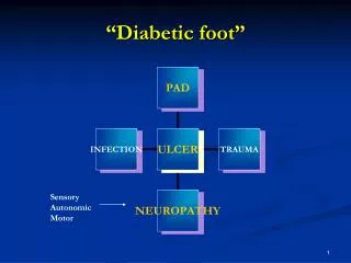
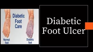
Pathophysiology Diabetic foot ulcers may have multiple causes Peripheral neuropathy (nerve damage) Peripheral vascular disease (poor pedal blood supply) Trauma Acute: any injury to the foot such as burns or cuts Chronic: due to foot deformities (changes of foot shape that lead to ill-fitting shoes and, thereby, ulceration)
Neuropathy Neuropathy Motor Sensory Autonomic Muscle wasting Foot weakness Postural deviation ↓ Proprioception, Unawareness of foot position Reduced sweating Stress on bones & joints Plantar pressure Dry skin ↓ nociception Fissures and cracks Callus formation Deformities, stress and shear pressures Trauma Ulcer Infection *Shunts: blood vessels that bypass capillaries and lead directly from arteries to veins
Ischaemic toes due to artherosclerosis Peripheral Arterial Disease Peripheral arterial disease Artherosclerosis narrows or blocks the arterial lumen Artheroma plaque narrowing the arterial lumen Foot ischaemia Foot ulcer Necrosis/ Gangrene Infection
Assestment The aim of the assessment is to examine each pathological cause that creates ulcers: 1) peripheral neuropathy peripheral arterial disease structural
How to Perform Proper Foot Examination Skin changes Evidence of infection Callous or ulcer Range of motion Charcot foot Structural Abnormalities Temperature Skin changes Ankle Brachial Index Peripheral Arterial Assessment Neuropathy Assessment 10 gram monofilament Tuning Fork (vibration)
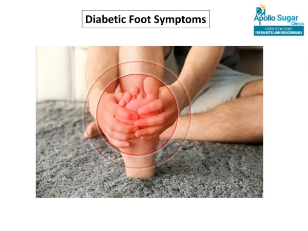
Assessment Peripheral Neuropathy History • burning, tingling, numbness of the foot Examination • Test for reduced power and reflexes that are evidence of muscular motor deficits. • Testsensationby skin pinprick (spinothalamic tracts), proprioception and vibration (dorsal columns)
AssessmentMonofilament for pressure sensation (pinprick sense) Place a 10g nylon Semmes-Weinstein monofilament to the skin Apply pressure until the monofilament buckles Inability to perceive the 10g of force applied by the monofilament is associated with clinically significant large fibre neuropathy and an increased risk of ulceration (sensitivity of 66 to 91%) Test 4 plantar sites on the forefoot (great toe and the base of 1st, 3rd and 5th metatarsals ) to identify 90% of patients with an insensate foot. Monofilament test
Assessment Tuning Fork (vibration) Apply a vibrating 128 Hz tuning fork to the bony prominence of the big toe If the patient cannot feel the vibration, gradually move the fork upwards Tuning fork test
AssessmentPeripheral Vascular Disease (PVD) History : claudication (calf pain after walking a specific distance) that is relieved by rest. However this is uncommon in people with diabetes due the concomitant neuropathy. Examination: Palpate the foot for temperature (cool in PVD); palpate the dorsalispedis pulse and, if absent, the posterior tibial pulse. Palpation of the dorsalis pedis pulse Palpation of the posterior tibial pulse
AssessmentInvestigations: ankle brachial pressure index Measure the blood pressure (BP) in the arm using a sphygmanometer Measure the blood pressure in the foot. Place a BP cuff around the calf and detect the dorsalispedis pulse using a small hand-held doppler. Inflate the cuff and slowly deflate until the pulse appears. The ankle brachial pressure index (ABPI) is the ratio of the ankle systolic pressure to brachial systolic pressure. ABPI is usually >1 but in the presence of peripheral vascular disease is <1. Doppler being used to detect the dorsalis pedis pulse
AssessmentStructural Abnormalities and Deformities Callus on plantar surface Structural abnormalities and deformities lead to bony prominences which are associated with high mechanical pressure on the overlying skin. Common abnormalities / deformities include: Callus Claw toes Charcot foot Nail deformities Nail deformity Charcot foot deformity Claw toes
Infected Ulcers • Signs suggesting infection include; • purulent secretions • presence of friable tissue (slugh) • foul odour The presence of infection needs to be defined clinically rather than microbiologically. An infected ulcer
AssessmentInfected Ulcers: Investigations Simple investigations include: • Tissue specimens or material obtained from the bottom of a wound for gram staining and culture for microbial sensitivity. • Full blood count, urea and electrolytes, inflammatory markers (ESR and CRP) for assessing severity of infection • Plain X-ray of the leg for signs of bone damage, presence of foreign body, or gas in soft tissue (gas gangrene)
Management Treatment of diabetic foot ulcers largely depends on the underlying causes: ischaemia, neuropathy or a combination of both.
Multidisciplinary Team Approach Wound care Pressure offloading Debridement (nonischemic wounds) Revascularisation Local factors Glycemic control Treat infection Address lower-extremity vascular status Systemic factors
ManagementUlcers due to Ischaemia • Medical: reduce cardiovascular risk factors • Surgical: revascularisation • Angioplasty • Open bypass surgery Ischaemic necrosis of a toe and an extensive plantar ulcer
ManagementUlcers due to Neuropathy The key to treatment here is to redistribute plantar pressure. Padding crutches, wheelchairs The common site for a neuropathic ulcer
ManagementWound Debridement • Debridement is the removal of necrotic and dead tissue in order to enhance healing. • Debridement is undertaken to: • Remove callus in neuropathic foot to lower plantar pressure • Assess the true dimension of the ulcer • Drain exudate and remove dead tissue • Take a deep swab for culture • Encourage healing and restore a chronic wound to an acute wound In both isacheamic and neuropathic ulcers, treatment is based on debridement of the wound and dressing application. Forcep and a scalpelis the usual technique by cutting away of all slough and non-viable tissue.
ManagementAmputation Amputation is made on clinical findings that the ulceration is not healing/ infection worsening in spite of intensive antibiotic therapy Signs include: • Extensive tissue loss • Unreconstructableischaemia • Failed revascularisation • Charcot’s of ankle with instability
ManagementInfected Ulcers - Antibiotics Empirically based upon clinical experience and local preferences Antibiotics are modified on the basis of clinical response and and wound culture / sensitivity results. For mild infections, 7-10 day course is usually sufficient. Severe infections may need up to 2-3 weeks of treatment.
Summary • Diabetic foot ulcers may have multiple causes • The aim of the assessment is to examine each pathological cause that creates ulcers • Multi-disciplinary approach needed • Treatment of diabetic foot ulcers largely depends on the underlying causes