Download

1 / 48
510 likes | 850 Views
Urinary System L 1 Functional Structures of the kidney. Prof. Madaya Dr Than Kyaw 1 October 2012. Urinary System . Waste products of metabolism – toxic (CO2, ammonia, etc) Removal from tissues – blood and lymph - respiratory system - skin – sweat & sebaceous gland

E N D
Urinary System L 1 Functional Structures of the kidney Prof. MadayaDr Than Kyaw 1 October 2012
Urinary System Waste products of metabolism – toxic (CO2, ammonia, etc) Removal from tissues – blood and lymph - respiratory system - skin – sweat & sebaceous gland Urinary system – Not only important for - removal of metabolic wastes - e.g. nitrogenous waste But also maintenance of body fluid and electrolyte balance (fluid homeostasis) - also has endocrine functions
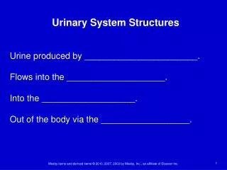
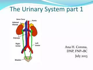
Urinary System • The urinary system consists of • paired kidneys and ureters, • a urinary bladder, • sphincter muscles • - and a urethra
Location of kidneys in animals Small ruminant
Kidneys • Paired organs suspended from the dorsal abdominal wall by a peritoneal fold and blood vessels that serve them • Located slightly cranial to the mid lumber region • Retroperitoneal structure – separated from the abdominal cavity by their envelopment of peritoneum • Renal artery and veins carry blood to and from the kidneys • Renal artery – arises directly from the aorta • Renal vein – empties directly into the caudal vena cava
Ventral view (dog) of kidneys showing renal arteries, veins and ureters and their portions relative to the aorta, vena cava and adrenal glands.
Kidneys Renal hilus – concave edge of the kidney – ureter, blood vessels, nerves and lymphatics enter or leave Neural innervation -- Sympathetic (adrenergic) division of autonomic nervous system; postganglionic renal nerves enter the hilus of kidney in association with the renal vessels -- Innervation to - renal vasculature, - all segments of the nephron, and - juxtaglomerular glandular cells
Types of kidney A. Heart-shaped - Horse B. Lobulated - Cattle C. Bean-shaped - most of the domestic animals (dog, cat, sheep) • Ventral views of right kidney • 1. Renal artery • 2. Renal vein • 3.ureter
Types of kidney (sagittal section) Unilobar kidney - carnivores, equine and small ruminants Multilobar - large ruminants, smooth surface, clear demarcation Multilobar - pig, cortical portion of the lobes fused
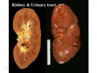
Medulla and cortex • Each kidney - composed of outer cortex and inner medulla • Pelvis • Hilus • Greater detail gross structures • Bisected preserved • sheep kidney
Medulla and cortex Red arrow = renal artery Blue arrow = renal vein 3 = Ureter 4 = Major calyx 5 = Minor calyx P = renal pyramid B = renal column Medulla - striations - Loops of Henle - Collecting tubules
Renal blood flow • At hilus – renal artery branches into smaller segmental arteries to supply sectors or segments of the mass of kidney tissues • Each segmental artery – divides to create lobar arteries which divide to yield interlober arteries that pass between the pyramids of the medulla • These interlobarartries branch into the arcuate arteries that arch over the base of renal pyramids. • Small interlobular arteries radiate outwards to supply the tissues of the cortex • 90% of blood entering the kidney supplies the cortical tissues where the bulk of nephrons are located • The veins trace the same pathways in reverse
Renal blood flow Aorta and renal artery
Renal blood flow INA = Interlobular artery AR = arcuate arteries IA = interlobar SA = Segmental artries
Distal convoluted tubules The nephron(Model) Renal glomeruli - yellow arrows Proximal convoluted tubule Descending loop of Henle Ascending loop of Henle CD = collecting duct Note: thin portion of ascending and descending loops
The nephron • Functional unit of the kidney • Understanding the function of nephron – essential for understanding kidney function • Nephron number – vary considerably among species • Within species – nephron number relatively constant Dogs - kidneys of large breed contain approximately similar numbers of nephrons as in small breed - compensation by having larger nephrons rather than more nephrons
The approximate number of nephrons in domestic animals and man
Types of nephron Identified by - location of glomeruli and - depth of penetration of loop of Henle into the medulla 2 types 1. Cortical or corticomedullarynephrons - glomeruli in the outer and middle cortices - loop of Henle extend to the junction of the cortex and medulla or into the outer zone of the medulla 2. Juxtamedullarynephrons - glomeruli in the cortex close to the medulla - loop of Henle extend more deeply into the medulla - some extend as deep as the renal pelvis
Types of nepharon Justamedullarynephrons - develop and maintain osmotic gradient from low to high in the outer medulla to the inner medulla respectively - 3% in pigs - 100% in cat - 14% in man Tubular fluid from both types of nephrons - enter collecting tubules and collecting ducts
Nephron components Glomerulus - tuft of capillaries - filtration function - afferent arteriole – conduct blood to the glomerulus - efferent arteriole – conduct blood away from glomerulus - efferent arterioles – peritubular capillaries – to vasarecti – - - to pelvis (see renal blood flow slide)
Nephron tubules and ducts (filtrate/fluid flow) Bowman capsule Proximal tubule Loop ofHenle Filtrate from glomerulus Distal tubule Collecting Duct Cortical collecting tubule Pelvis Ureter Bladder (store) urethra
Functional nephron with blood supply 1. Bowman capsule 2. Proximal tubule 3. Descending limb of LOH 4. Thin ascending limb of LOH 5. Thick ascending limb of LOH 6. Distal tubule 7. Connecting tubule 8. Cortical collecting tubule 9. Outer medullary collecting duct 10. Inner medullary collecting duct 11. Afferent arteriole 12. Glomeruls 13. Efferent arteriole Periyubular capillaries Vasa recta To renal vein
Loop of Henle Composed of 3 segments • Thin descending limb • Thin ascendind limb • Thick ascending limb • Lumen diameter does not change • Descending limb of cortical nephrons - only go as deep as the outer space of the outer medulla • Descending limb of juxtamedullarynephrons - may extend up to the pelvis
Juxtaglomerularappatatus (JG apparatus) JG apparatus - The junction of distal tubules and glomerulus - JG cells + macula densa + extraglomerulamesangial cells - regulate amount of blood flowing to the kidney - secretion of enzyme renin – important for the formation of angiotensin II Macula densa - collectively named tubular cells involved in the JG apparatus Juxtaglomerular (JG) cells – cells enclosing afferent and efferent arterioles in the junction of distal tubule and glomerulus Mesangial cells – cells between macula densa and arterioles
Formation of Urine Terminology Renal Blood Flow (RBF) - the rate at which blood flows to the kidney (in ml per minute) Renal Plasma Flow (RPF) - refers to part of RBF that is plasma Glomerular Filtration Rate (GFR) - the rate at which filtrate is formed (in ml per min) Filtrate fraction (FF) - the ratio of GFR to RPF (GFR:RPF)
Some approximate value of renal function variables(11.35 kg dog in a normal state of hydration)
Formation of Urine GFR – normally about 100 times that of RBF - 3-5 ml/kg BW/min High GFR - allows a continuous filtration of plasma - rapid removal of toxic substances from the body - if they can readily pass through the glomerular filtration barrier and not reabsorbed from the renal tubules
Formation of Urine 3 processes involved in the urine formation Glomerular filtration Tubular reabsorption (selective) Tubular secretion (selective)
Glomerular filtration • Kidneys have functional counter part of 2 capillary beds - glomerular capillaries (filter) - peritubular capillaries (reabsorption and/or secretion) • Glomeruli - Have high pressure system (high hydrostatic pressure) - favour filtration • Peritubular - Low pressure system - favourreabsorption
High pressure system in the glomerulusfavour net formation of filtrate (fluid) in the capsular space As filtrate flows away from the capsule – colloidal osmotic pressure may be negligible COP = colloidal osmotic pressure HP = hydrostatic pressure Net filtration pressure = 60 – (18+32) = 10 mm Hg
Glomerular filtration • Blood flows through glomeruli – large quantity of filtrate formed • k/s glomerular filtrate Physical barriers for filtration 1. capillary endothelium of the glomerulus 2. inner layer of Bowman’s capsule 3. basement membrane between endothelium and glomerulus
Glomerular filtration Glomerular endothelium - fenestrated – porous - highly permeable
PP = pedicels of podocyte FS = filtration slit US = filtrate or urinary space E = endothelial pore EP = endothelial pores P = podocytes GBM = glomerular basement membreane
Glomerular filtration • Podocytes • Cells of the inner layer of Bowmen’s capsule • Have cellular extensions that rest on the glomerular basement membrane • Slit-like pores between the extensions permit the passage of the filtrate
Glomerular filtration Glomerular filtration barrier - acts like a sieve - substances up to a molecular weight of 65,000 pass through the barrier - small percentage of plasma proteins pass through it - glucose, amino acids, urea, creatinine, Na, K, chlorine, and bicarbonate ions – readily cross the barrier - concentration in the initial filtrate – about same as plasma
Glomerular filtration Pressure maintained by vasoconstriction of efferent vessels Porous walls + high pressure Bowman’s capsule Water and solutes <10kDa out Water, sugars, salts, amino acids, Urea (sometimes assisted by active transport) Primary Urine: Dilute, no proteins etc. Large things (e.g. proteins) remain behind
Glomerular filtration Proteinuria - presence of abnormal amounts of protein voided in the urine - kidney diseases that localize in or primarily affect glomeruli - associated with proteinuria or hematuria
Factors controlling Glomerular filtration Rate Forces determining rate of movement of fluid across the glomerular filtration barrier are generally similar as those that determine fluid movement out of capillaries throughout the body. Diameter of afferent Dilatation of arterioles - increase blood flow to glomeruli - increase HP and potential for filtration Constriction of efferent arterioles - increase glomerular HP
Factors controlling Glomerular filtration Rate For a given molecular size - positively charges molecules are more readily filtered than negatively charged molecules - this is because of negatively charged (anionic) sites in the glomerular basement membrane - normally most of plasma proteins are restricted from filtration - kidney diseases - change of electrostatic charge on the glomerular membrane – allow filtration
Factors controlling Glomerular filtration Rate Effective filtration pressure - the pressure tending to force fluid out of the capillaries - It is the difference between the blood hydrostatic pressure in the capillary and osmotic pressure generated by plasma protein of the blood in the capillary - also hydrostatic pressure and osmotic pressure of urinary space of Bowman’s capsule - important, especially in diseased state (blockage of urinary tract or renal tubules)
Renin-angiotensin system Angiotensin converting enzyme (Vascular endothelium) Renin - secreted by juxtaglomerula cells of the kidney Circulating blood globulin, angiotensinogen to form Angiotensin I Angiotensin II Low blood pressure Aldosterone secretion (Zonaglomerulosa) - Systemic arteriolar vasoconstriction - Increase systemic blood pressure Promote Na reabsorption And retention of water