Download
1 / 11
120 likes | 259 Views
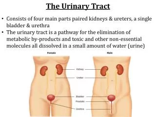
Pathology of Kidney and the Urinary tract . Dr. Amar C. Al- Rikabi Dr. Hala Kasouf Kfouri. Normal Kidney, Congenital and Cystic Renal Diseases and Acute Renal Failure . Lecture -1: .

E N D
Pathology of Kidney and the Urinary tract Dr. Amar C. Al-Rikabi Dr. HalaKasoufKfouri
Normal Kidney, Congenital and Cystic Renal Diseases and Acute Renal Failure Lecture -1:
This paraffin embedded 2µm section illustrates a normal glomerulus with normal vascular pole with minimal periglomerular interstitial fibrosis and surrounding intact tubules
This glomerulus shows only minimal abnormalities by electron microscopy, with rare Vacuoles and blebs in the podocytes.
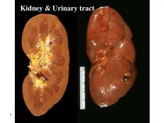
Bisected kidney shows a less severe form of CRD with multiple cysts and focally a significant degree of disorganization of parenchymal architecture.
Autosomal recessive (infantile) polycystic disease (ARPKD). Elongated streaks represent dilated tubules.
Histology of ADPKD shows a single glomerulus alongside several cysts Cut surface of ADPKD showing variable-size, irregular cysts with no recognizable intervening normal parenchyma. Most cysts contain clear fluid and the pelvis is distorted.
Late stage of ADPKD showing greatly enlarged renal profiles due to innumerable cysts of varying size replacing the renal parenchyma.
Elongated, stretched out, regenerating proximal tubular lining cells encompass the necrotic epithelium. Kidney in acute tubular necrosis (ATN) showing pale, swollen cortex and congested medulla.