Download
1 / 1
10 likes | 152 Views
Fiber Demixing with the Tensor Distribution Function avoids errors in Fractional Anisotropy maps. Liang Zhan 1 ,Alex D. Leow 2,3 , Neda Jahanshad 1 , Arthur W. Toga 1 , Paul M. Thompson 1. 1 Laboratory of Neuro Imaging, Dept. of Neurology, UCLA School of Medicine, Los Angeles, CA, USA

E N D
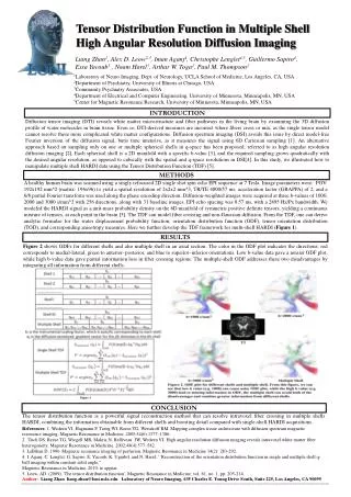
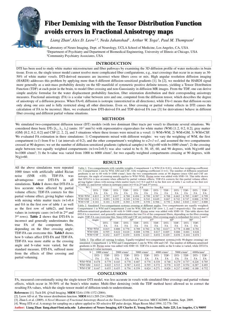
Fiber Demixing with the Tensor Distribution Function avoids errors in Fractional Anisotropy maps Liang Zhan1,Alex D. Leow2,3, Neda Jahanshad1 , Arthur W. Toga1, Paul M. Thompson1 1Laboratory of Neuro Imaging, Dept. of Neurology, UCLA School of Medicine, Los Angeles, CA, USA 2Department of Psychiatry and Department of Biomedical Engineering, University of Illinois at Chicago, USA 3Community Psychiatry Associates, USA INTRODUCTION DTI has been used to study white matter microstructure and fiber pathways by examining the 3D diffusion profile of water molecules in brain tissue. Even so, the single tensor model cannot resolve more complicated fiber configurations, e.g., tract crossings that occur in as many as 30-50% of white matter voxels. DTI-derived measures are incorrect where fibers cross or mix. High angular resolution diffusion imaging (HARDI) addresses this problem by applying more than 6 different diffusion-sensitized gradients [1]. In [2], we modeled the HARDI signal more generally as a unit-mass probability density on the 6D manifold of symmetric positive definite tensors, yielding a Tensor Distribution Function (TDF) at each point in the brain, to model fiber crossing and non-Gaussianity in diffusion MR images. From the TDF, one can derive simple analytic formulae for the water displacement probability function, fiber orientation distribution and their corresponding anisotropy measures. Fractional anisotropy (FA) is a scalar value between zero and one, computed from the diffusion tensor, which describes the degree of anisotropy of a diffusion process. When FA=0, diffusion is isotropic (unrestricted in all directions), while FA=1 means that diffusion occurs only along one axis and is fully restricted along all other directions. Even so, fiber crossing or partial volume effects in DTI causes the calculation of FA to be incorrect. Here, we evaluated how DTI-derived FA and TDF-derived FA (see [3] for derivations) behave in different fiber crossing and different partial volume situations. METHODS We simulated two-compartment diffusion tensor (DT) models (with two dominant fiber tracts per voxel) to illustrate several situations. We considered three basic DTs [λ1, λ2, λ3] (units: 10-3 mm2/s) with representative eigenvalues for white matter (WM) [1.2, 0.2, 0.2], gray matter (GM) [0.2, 0.2, 0.2] and CSF [2, 2, 2], and 3 situations where these tissues were mixed in a voxel: 1) WM+WM, 2) WM+GM, 3) WM+CSF. We evaluated FA estimation in three simulations: 1) Compartments mixed with different weights: we vary the weighting of WM, the first compartment (w1) from 0 to 1 (at intervals of 0.2), and the other compartment’s weighting to w2=1-w1, and assumed the two compartments crossed at 90 degrees; we set the number of diffusion-sensitized gradients (spherical samples) to N(g)=60 with b=1000 s/mm2; 2) the crossing angle between two equally weighted compartments (w1=w2=0.5) was also varied to be 0, 30, 45, 60, and 90 degrees, with N(g)=60 and b=1000 s/mm2; 3) the b-value was varied from 1000 to 6000 s/mm2, for two equally-weighted compartments crossing at 90 degrees, with N(g)=60. RESULTS All the above simulations were repeated 1000 times with artificially added Rician noise (SNR =10). TDF-FA was advantageous over DTI-FA in all simulations. Table 1 shows that DTI-FA is less accurate when affected by partial volume effects; TDF-FA corrects for this partial volume effect, giving higher values with mixing white matter tracts (w1=0.4 and 0.6 in the first row of table 1 as well as the first row of table2), and lower values in isotropic cases (w1=0 in 2nd and 3rd rows). Table 2 shows that DTI-FA is incorrect and generally understimates the true FA of the component fibers, depending on the fiber crossing angle; TDF-FA can overcome this. Table3 shows how b values affect DTI-FA and TDF-FA. TDF-FA was more stable as the crossing angle and b-value were varied, but the standard measure, DTI-FA, suffered more from the effects of fiber crossing and partial voluming. CONCLUSION FA, measured conventionally using the single-tensor DTI model, was less accurate in voxels with simulated fiber crossings and partial volume effects, which occur in 30-50% of the brain’s white matter. Multi-fiber demixing (with the TDF method here) allowed us to correct the resulting FA values, which the single-tensor model of diffusion tends to underestimate. References: [1]. Tuch DS. Q-ball Imaging. MRM 52(6):1358-1372 (2004). [2]. Leow AD et al. The tensor distribution function. MRM 61(1):205-214 (2008) [3]. Zhan L et al.(2009). A Novel Measure of Fractional Anisotropy Based on the Tensor Distribution Function. MICCAI2009, London, Sept. 2009. [4]. Wong STS et al. A strategy for sampling on a sphere applied to 3D selective RF pulse design. Magn Reson Med 1994; 32:778–784. Author: Liang Zhan liang.zhan@loni.ucla.edu Laboratory of Neuro Imaging, 635 Charles E. Young Drive South, Suite 225, Los Angeles, CA 90095