Download
1 / 7
90 likes | 215 Views
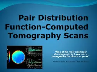
The Atomic Pair Distribution Function Method. Getting to know your atomic neighborhood. Inroduction. Modern materials are often disordered. Standard crystallographic methods lose the aperiodic (disorder) information. We would like to be able to sit on an atom and look at our neighborhood.

E N D
The AtomicPair Distribution Function Method Getting to know your atomic neighborhood
Inroduction • Modern materials are often disordered. • Standard crystallographic methods lose the aperiodic (disorder) information. • We would like to be able to sit on an atom and look at our neighborhood. • The PDF method allows us to do that (see next slide): • First we do a neutron or x-ray diffraction experiment • Then we correct the data for experimental effects • Then we Fourier transform the data to real-space
Obtaining the PDF Structure function Raw data PDF
What is the PDF? • (a) The red ball is a C60 molecule. C60 forms a solid by the molecules clustering (b). The scattering and PDF are shown in (c) and (d) respectively. • Sit on an atom and look at your neighborhood. The nearest neighbor is at 1.4A distance, the second neighbor at 2.2A and so on. There are sharp peaks in G(r) at these positions. This is the structural information in the PDF. • There are no sharp peaks beyond 7.1A, the diameter of the ball because the balls are spinning with respect to each other. The PDF can see this.
Full Profile PDF fitting using PDFFIT • We extract information from the PDF by fitting structural models to the data • We use Full-profile least-squares fitting of the PDF using the program PDFfit • The red line is the PDF from the model, the blue line the data, the green line the difference. • The data are neutron data from LaMnO3 collected 10K at IPNS, Argonne National Laboratory, IL. • Ref: Proffen et al., Phys. Rev. B 60, 9973 (1999).
r1 << x r1 r2 ~ x/2 r2 Intra-domain structure Inter-domain structure Observing Domains in the PDF The PDF gives different information on different length-scales. We can see the structure within a domain at low-r and between domains at high-r.
Goodbye • Please look elsewhere on the web-page, or check our publication list, to see the ways we are applying the PDF to learn about materials. • Thanks for the visit!