Download
1 / 82
830 likes | 1.54k Views
ARRHYTHMIA Edited by Yingmin Chen. Definition of Arrhythmia: The Origin, Rate, Rhythm, Conduct velocity and sequence of heart activation are abnormally . Anatomy of the conducting system. Pathogenesis and Inducement of Arrhythmia . Some physical condition Pathological heart disease

E N D
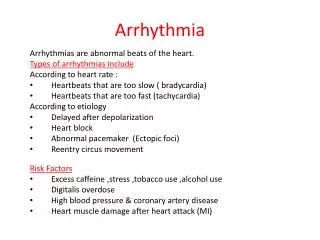
Definition of Arrhythmia: The Origin, Rate, Rhythm, Conduct velocity and sequence of heart activation are abnormally.
Pathogenesis and Inducement of Arrhythmia • Some physical condition • Pathological heart disease • Other system disease • Electrolyte disturbance and acid-base imbalance • Physical and chemical factors or toxicosis
Mechanism of Arrhythmia • Abnormal heart pulse formation • Sinus pulse • Ectopic pulse • Triggered activity • Abnormal heart pulse conduction • Reentry • Conduct block
Classification of Arrhythmia • Abnormal heart pulse formation • Sinus arrhythmia • Atrial arrhythmia • Atrioventricular junctional arrhythmia • Ventricular arrhythmia • Abnormal heart pulse conduction • Sinus-atrial block • Intra-atrial block • Atrio-ventricular block • Intra-ventricular block • Abnormal heart pulse formation and conduction
Diagnosis of Arrhythmia • Medical history • Physical examination • Laboratory test
Therapy Principal • Pathogenesis therapy • Stop the arrhythmia immediately if the hemodynamic was unstable • Individual therapy
Anti-arrhythmia Agents • Anti-tachycardia agents • Anti-bradycardia agents
Anti-tachycardia agents • Modified Vaugham Williams classification • I class: Natrium channel blocker • II class: ß-receptor blocker • III class: Potassium channel blocker • IV class: Calcium channel blocker • Others: Adenosine, Digital
Anti-bradycardia agents • ß-adrenic receptor activator • M-cholinergic receptor blocker • Non-specific activator
Clinical usage Anti-tachycardia agents: • Ia class: Less use in clinic • Guinidine • Procainamide • Disopyramide: Side effect: like M-cholinergic receptor blocker
Anti-tachycardia agents: • Ib class: Perfect to ventricular tachyarrhythmia 1. Lidocaine 2. Mexiletine
Anti-tachycardia agents: • Ic class: Can be used in ventricular and/or supra-ventricular tachycardia and extrasystole. 1. Moricizine 2. Propafenone
Anti-tachycardia agents: • II class: ß-receptor blocker • Propranolol: Non-selective • Metoprolol: Selective ß1-receptor blocker, Perfect to hypertension and coronary artery disease patients associated with tachyarrhythmia.
Anti-tachycardia agents: • III class: Potassium channel blocker, extend-spectrum anti-arrhythmia agent. • Amioarone: Perfect to coronary artery disease and heart failure patients • Sotalol: Has ß-blocker effect • Bretylium
Anti-tachycardia agents: • IV class: be used in supraventricular tachycardia • Verapamil • Diltiazem • Others: Adenosine: be used in supraventricular tachycardia
Anti-bradycardia agents • Isoprenaline • Epinephrine • Atropine • Aminophylline
Proarrhythmia effect of antiarrhythmia agents • Ia, Ic class: Prolong QT interval, will cause VT or VF in coronary artery disease and heart failure patients • III class: Like Ia, Ic class agents • II, IV class: Bradycardia
Non-drug therapy • Cardioversion: For tachycardia especially hemodynamic unstable patient • Radiofrequency catheter ablation (RFCA): For those tachycardia patients (SVT, VT, AF, AFL) • Artificial cardiac pacing: For bradycardia, heart failure and malignant ventricular arrhythmia patients.
Sinus tachycardia • Sinus rate > 100 beats/min (100-180) • Causes: • Some physical condition: exercise, anxiety, exciting, alcohol, coffee • Some disease: fever, hyperthyroidism, anemia, myocarditis • Some drugs: Atropine, Isoprenaline • Needn’t therapy
SinusBradycardia • Sinus rate < 60 beats/min • Normal variant in many normal and older people • Causes: Trained athletes, during sleep, drugs (ß-blocker) , Hypothyriodism, CAD or SSS • Symptoms: • Most patients have no symptoms. • Severe bradycardia may cause dizziness, fatigue, palpitation, even syncope. • Needn’t specific therapy, If the patient has severe symptoms, planted an pacemaker may be needed.
Sinus Arrest or Sinus Standstill • Sinus arrest or standstill is recognized by a pause in the sinus rhythm. • Causes: myocardial ischemia, hypoxia, hyperkalemia, higher intracranial pressure, sinus node degeneration and some drugs (digitalis, ß-blocks). • Symptoms: dizziness, amaurosis, syncope • Therapy is same to SSS
Sinoatrial exit block (SAB) • SAB: Sinus pulse was blocked so it couldn’t active the atrium. • Causes: CAD, Myopathy, Myocarditis, digitalis toxicity, et al. • Symptoms: dizziness, fatigue, syncope • Therapy is same to SSS
Sinoatrial exit block (SAB) • Divided into three types: Type I, II, III • Only type II SAB can be recognized by EKG.
Sick Sinus Syndrome (SSS) • SSS: The function of sinus node was degenerated. SSS encompasses both disordered SA node automaticity and SA conduction. • Causes: CAD, SAN degeneration, myopathy, connective tissue disease, metabolic disease, tumor, trauma and congenital disease. • With marked sinus bradycardia, sinus arrest, sinus exit block or junctional escape rhythms • Bradycardia-tachycardia syndrome
Sick Sinus Syndrome (SSS) • EKG Recognition: • Sinus bradycardia, ≤40 bpm; • Sinus arrest > 3s • Type II SAB • Nonsinus tachyarrhythmia ( SVT, AF or Af). • SNRT > 1530ms, SNRTc > 525ms • Instinct heart rate < 80bmp
Sick Sinus Syndrome (SSS) • Therapy: • Treat the etiology • Treat with drugs: anti-bradycardia agents, the effect of drug therapy is not good. • Artificial cardiac pacing.
Premature contractions • The term “premature contractions” are used to describe non sinus beats. • Common arrhythmia • The morbidity rate is 3-5%
Atrial premature contractions (APCs) • APCs arising from somewhere in either the left or the right atrium. • Causes: rheumatic heart disease, CAD, hypertension, hyperthyroidism, hypokalemia • Symptoms: many patients have no symptom, some have palpitation, chest incomfortable. • Therapy: Needn’t therapy in the patients without heart disease. Can be treated with ß-blocker, propafenone, moricizine or verapamil.
Atrial tachycardia • Classify by automatic atrial tachycardia (AAT); intra-atrial reentrant atrial tachycardia (IART); chaotic atrial tachycardia (CAT). • Etiology: atrial enlargement, MI; chronic obstructive pulmonary disease; drinking; metabolic disturbance; digitalis toxicity; electrolytic disturbance.
Atrial tachycardia • May occur transient; intermittent; or persistent. • Symptoms: palpitation; chest uncomfortable, tachycardia may induce myopathy. • Auscultation: the first heart sound is variable
Intra-atrial reentry tachycardia (IART) • ECG characters: • Atrial rate is around 130-150bpm; • P’ wave is different from sinus P wave; • P’-R interval ≥ 0.12” • Often appear type I or type II, 2:1 AV block; • EP study: atrial program pacing can induce and terminate tachycardia
Automatic atrial tachycardia (AAT) • ECG characters: • Atrial rate is around 100-200bpm; • Warmup phenomena • P’ wave is different from sinus P wave; • P’-R interval≥ 0.12” • Often appear type I or type II, 2:1 AV block; • EP study: Atrial program pacing can’t induce or terminate the tachycardia
Chaotic atrial tachycardia (CAT) • Also termed “Multifocal atrial tachycardia”. • Always occurs in COPD or CHF, • Have a high in-hospital mortality ( 25-56%). Death is caused by the severity of the underlying disease. • ECG characters: • Atrial rate is around 100-130bpm; • The morphologies P’ wave are more than 3 types. • P’-P’, P’-R and R-R interval are different. • Will progress to af in half the cases • EP study: Atrial program pacing can’t induce or terminate the tachycardia
Therapy • IRAT:Esophageal Pulsation Modulation, RFCA, Ic and IV class anti-tachycardia agents • AAT: Digoxin, IV, II, Ia and III class anti-tachycardia agents; RFCA • CAT: treat the underlying disease, verapamil or amiodarone. • Associated with SSS: Implant pace-maker.
Atrial flutter • Etiology: • It can occur in patients with normal atrial or with abnormal atrial. • It is seen in rheumatic heart disease (mitral or tricuspid valve disease), CAD, hypertension, hyperthyroidism, congenital heart disease, COPD. • Related to enlargement of the atria • Most AF have a reentry loop in right atrial
Atrial flutter • Symptoms: depend on underlying disease, ventricular rate, the patient is at rest or is exerting • With rapid ventricular rate: palpitation, dizziness, shortness of breath, weakness, faintness, syncope, may develop angina and CHF.
Atrial flutter • Therapy: • Treat the underlying disease • To restore sinus rhythm: Cardioversion,Esophageal Pulsation Modulation, RFCA, Drug (III, Ia, Ic class). • Control the ventricular rate: digitalis. CCB, ß-block • Anticoagulation
Atrial fibrillation • Subdivided into three types: paroxysmal, persistent, permanent. • Etiology: • Morbidity rate increase in older patients • Etiology just like atrial flutter • Idiopathic • Mechanism: • Multiple wavelet re-entry; • Rapid firing focus in pulmonary vein, vena cava or coronary sinus.
Atrial fibrillation • Manifestation: • Affected by underlying diseases, ventricular rate and heart function. • May develop embolism in left atrial. Have high incidence of stroke. • The heart rate, S1 and rhythm is irregularly irregular • If the heart rhythm is regular, should consider about (1) restore sinus rhythm; (2) AF with constant the ratio of AV conduction; (3) junctional or ventricular tachycardia; (4) slower ventricular rate may have complete AV block.
Atrial fibrillation • Therapy: • Treat the underlying disease • Restore sinus rhythm: Drug, Cardioversion, RFCA, Maze surgery • Rate control:digitalis. CCB, ß-block • Antithrombotic therapy: Aspirine, Warfarin