Download
1 / 33
360 likes | 965 Views
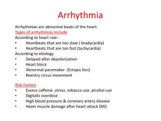
Arrhythmia. Arrhythmia. Arrhythmia prebared by Waleed Sammody Mohammad Mansour Supervised by Jehad Bani Aodih. Basic Facts An arrhythmia is an abnormality or disturbance in the rate or rhythm of the heartbeat.

E N D
Arrhythmia Arrhythmia
Arrhythmiaprebared by Waleed SammodyMohammad MansourSupervised byJehad Bani Aodih
Basic FactsAn arrhythmia is an abnormality or disturbance in the rate or rhythm of the heartbeat.
Arrhythmias are caused byproblems with the heart's electrical system, such as abnormal formation of the electrical impulses that begin heartbeats or by a disruption of the pattern of conduction of those impulses.
Because the ventricles are primarily responsible for moving blood through the body, ventricular arrhythmias are often more serious than other arrhythmias.
CAUSES AND RISK FACTORSCauses of ventricular arrhythmias include: 1- Abnormal electrolyte (mineral, such as potassium and magnesium) levels in the blood 2- Electrocution
3-Heart attack (myocardial infarction) 4-Unstable angina (chest pain). 5-Scarring resulting from a heart attack
Ventricular arrhythmias include Premature ventricular complexes (PVCs), which are premature heartbeats; Ventricular tachycardia, an abnormally fast heartbeat; and Ventricular fibrillation, in which the heart quivers rather than contracts. An electrocardiogram of an episode of sustained ventricular tachycardia.
Premature ventricular contraction (PVC): The ventricles fire an early impulse which causes the heart to beat earlier causing irregularity in the heart rhythm.
Ventricular Tachycardia • This is a dangerous type of rapid heart rhythm because it is usually associated with poor cardiac output (amount of blood ejected out of the heart) • It results from abnormal tissues in the ventricles generating a rapid and irregular heart rhythm.
Ventricular Tachycardia Rate Rhythm Ventricular > 100 bpm Regular Atrial None P-Wave Absent P-R Interval Absent QRS Complex > .10 seconds
Both ventricular tachycardia and ventricularfibrillation are considered lethal arrhythmias Only ventricular fibrillation, is linked to the clinical term “sudden death.” This rhythm is not able to support life and will lead to clinical death if untreated • Ventricular fibrillation All output from the heart stops, blood pressure falls rapidly, and the patient loses consciousness .
During ventricular fibrillation the heart is electrically stimulated by multiple ectopic sites so that instead of contracting rhythmically in one united wave of depolarization, the muscle actually fibrillates
WHAT ARE THE SYMPTOMS? Diminished or irregular pulse; Fatigue; Shortness of breath; Fainting (syncope); Low blood pressure; Chest pain; and Palpitations (awareness of one's own heartbeat); Cardiac arrest.
Ventricular fibrillation An electrocardiogram reflecting the irregular, pulseless electrical activity of ventricular fibrillation
TREATMENT APPROACH Many cases of arrhythmias may not require treatment. Other arrhythmias can be treated by treating any underlying heart disease. Treatments for ventricular arrhythmia includes
DefibrillationMedication (beta-blockers and antiarrhythmic agents); Radiofrequency catheter ablation; Angioplasty; and Pacemaker implantation.
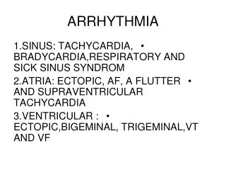
Heart Block Heart Block
Heart block, also called atrioventricular block or A-V block, is an abnormality of the spread or flow of electrical activity from the upper heart chambers, the atria, to the lower chambers of the heart, the ventricles.
Atrioventricular block (AVB): the sinus node may be generating heart beats causing the atria to contract at a normal rate, however not every electrical impulse coming from the atria is being passed down to the ventricles by the atrioventricular node due to a block in conduction. There are various types of AV block depending upon the mechanism of block.
First-degree heart block Heart beat impulses are delayed in the A-V node, but ultimately reach the ventricles. Sometimes, first-degree heart block can eventually lead to other forms of heart block. .
In first-degree heart block, the ECG shows one QRS wave for every P wave, but the pause is greater than normal
Second-degree heart block Heart beat impulses are delayed or blocked in or around the A-V node, and some of the impulses fail to reach the ventricles; Second-degree heart block is further divided into two sub-types: Type I second-degree heart block, also called Mobitz Type I heart block or the Wenckebach phenomenon. Type II second-degree heart block, also called Mobitz Type II block.
second degree block referred to as Mobitz I or Wenckebach • Occasionally sinus impulses will pass through the AV node at slower and slower rates until excitation is actually blockedAfter that the cycle of delay-delay-block repeats itself • This phenomenon is a form of second degree block referred to as Mobitz I or Wenckebach.
Mobitz II is another form of second-degree block • Mobitz II occurs within the context of a basic rhythm when a P wave occurs but is not followed by a QRS. This is a more serious form of second-degree block as it occurs without warning • The basic rhythm would be regular except for the periods of the block. These occur most often because there is block below the bundle of His The P to P interval is once again regular. The QRS response, because of the “dropped beat,” will appear irregular
In second-degree type II block, notice that the P wave (4th bump) isn't followed by the QRS wave, because the ventricles weren't activated.
Third-degree heart block Also called complete heart block, each sinus node impulse is completely interrupted in the A-V node or beyond, and the ventricles must generate their own impulse to contract. Depending on its cause, third-degree block may be transient (temporary) or permanent. When no impulses from the atria excite the ventricle, a situation of complete block exists
Because they are capable of self-automaticity, a ventricular rhythm is present • Clearly this is the most serious form of heart block as the ventricles are now left on their own to beat • However, at a rate inherent to the ventricles the patient’s rhythm will significantly slow • In addition, the effect of atrial kick is lost
THANK YOU thank you