Download
1 / 59
700 likes | 1.55k Views
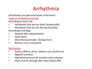
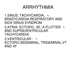
Arrhythmia. Classification. Abnormal origin ----sinus arrhythmia ----ectopic rhythm:passivity—escape ---premature contraction tachycardia

E N D
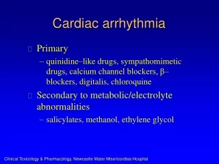
Classification Abnormal origin ----sinus arrhythmia ----ectopic rhythm:passivity—escape ---premature contraction tachycardia flutter and fibrillation Abnormal conduction ----physiological block: ----pathological block: S-AB; A-VB; LBBB; RBBB ----accessory pathway: pre-excitation syndrome
Electrophysiology • Automaticity • Excitability • Absolute refractory period (200ms) • Effective refractory period (210ms) • Ralative refractory period (50-100ms) • Conductivity
Sinus rhythm features : (1) Every P wave is following by a QRS complex; (2) P wave is upright in lead I, II, aVF, V4-V6, inverse in aVR; (3)P-R interval ≥ 0.12sec; (4)Normal rate is 60-100 beats/min
Sinus Bradycardia (1)Sinus rhythm (2)Heart rate <60bpm (R-R interval or P-P interval >1.0 sec )
Factors associated with sinus bradycardia (1) Physiologic Laborers and trained athletes Emotional states leading to syncope (2) Pathologic -blocker Hypothyroidism
Sinus Tachycardia (1)Sinus rhythm, rate > 100 bpm The R-R interval (or the P-P interval) <0.60 sec. (2) P-R and Q-T interval are shorter than usual (3) S-T segment is slight depression, T waves may be flattened
Factors associated with sinus tachycardia (1) Physiologic Exercise Strong emotion Anxiety states (2) Pathologic Fever Hemorrhage Anemia Myocarditis Hyperthyroidism
Sinus arrhythmia • Sinus rhythm and PR interval, • Difference of P--P interval > 0.12sec in the same lead
Sinus arrest The P wave missed for a short time
Sick Sinus Syndrome (SSS) (1)Sinus bradycardia (HR<50/min); (2)Sinus arrest or SA block; (3)Tachycardia:Atrial tachycardia, Atrial Flutter, Atrial fibrillation; (4) AV block.
1. Premature Ventricular Contraction (1) Ventricular complex (QRS) is not preceded by a premature P' wave. (2) Premature QRS complex is the wider and the bizarre , Duration of QRS> 0.12 sec. T wave in direction is opposite to QRS complex . (3) Complete compensatory pause [bɪˈzɑr]
ventricular premature beat Complete compensatory pause=(P-P)X 2 P P P X 2X
bigeminy trigeminy tetrageminy
2. Atrial Premature Contractions (1) The premature P' wave differs in contour from the normal P wave in the same lead. (2) The P'-R interval >0.12s. (3) There may be a noncompensatory pause.
Premature atrial contractions.A, PAC after the fourth sinus beat.B, blocking of the PAC occurs after the fourth sinus beat. The premature P waves falls on the T wave of the preceding beat and is not followed by a QRS complex.
3. Premature junctional contraction (1) A premature normal-appearing QRS complex. (2) The junctional P wave (P’) may be appear before, in, and after the QRS. (3) Usually a complete compensatory pause.
Tachycardia Reentry Requires: Two conducting pathways Unidirectional block in one Slow conduction in the other
1. Paroxysmal supraventricular tachycardia (PSVT) a. Heart rate between 160 – 250 bpm. b. A precisely regular rhythm with normal QRS.
2. Ventricular Tachycardia a)The rate is 140200/min and the rhythm is very slightly irregular. b)QRS complex is the wider and the bizarre , Duration of QRS >0.12 sec. c)P wave dissociated from QRS; The rate of P wave is less than The rate of QRS d) Ventricular capture ; e)Fusion beats are present.
3.Nonparoxysmal Tachycardia • Nonparoxysmal junctional Tachycardia, The heart rate is 70130/min • Nonparoxysmal ventricular Tachycardia. The heart rate is 60100/min
1. Atrial Flutter (1)Absence of normal P waves; (2)P waves replaced by saw-tooth flutter wave (F waves); (3)Flutter waves seen best in leads II, III,aVF; (4)F waves always uniform in size, shape and frequency and absence of isoelectric line between F waves; (5)Regular atrial rhythm with a rate of 250-350 /min; (6)Ventricular response of 1:1,2:1,3:1,4:1 or higher
2. Atrial Fibrillation (1)Absence of clear P waves ; (2)P waves replaced by f waves; (3)f waves: irregular in size, shape, best seen in lead V1; (4)Rate of f waves is 350 - 600/min ; (5)Irregularly irregular ventricular rate; (6)Generally, duration of QRS complex <0.12sec;
Ventricular Flutter and Ventricular fibrillation Ventricular flutter: It is impossible to separate the QRS complexes from the ST segment and the T waves Ventricular fibrillation: The ECG shows fine or coarse waves that are rapid, and irregular in size, shape, and width .
1. First Degree A-V Block • Prolonged P-R interval: P-R interval > 0.20sec. in adults (varies with heart rate)
2.Second Degree A-V Block • (1) Mobitz type I (Wenckebach phenomenon). • The pattern is a progressive prolongation of the P-R interval until a beat is dropped. • The first beat after the pause has the shortest P-R interval, which may or may not be normal.
(2) Mobitz type II • There is a fixed numerical relationship between atrial and ventricular impulses, which may be 2:1 (2 atrial beats to one ventricular beat) or 3:1 or 4:1.
Third Degree A-V Block (Complete heart block) (1) The atrial and the ventricular rhythms are absolutely, independent of one another. (There is no relationship of P to QRS.) (2) atrial rate > ventricular rate. • QRS is 0.12 sec. or greater.
4. Complete Right Bundle Branch Block (1) Right axis deviation. (2) QRS≥0.12 sec. (3) rsR’ pattern (M pattern ) in V1 or V2; (4) Wide and slurred S wave in leads 1, V5 and V6 . (5) ST-T changes in leads V1 and V2 .