Download
1 / 3
30 likes | 233 Views
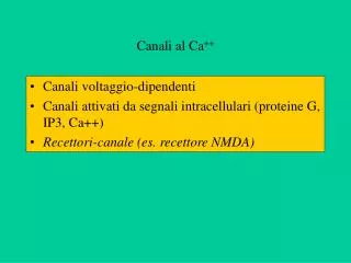
長期給予嗎啡或安非他命對大白鼠或幼鼠腦部興奮性的表現與 NMDA 受體所產生的影響.

E N D
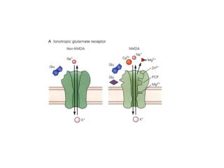
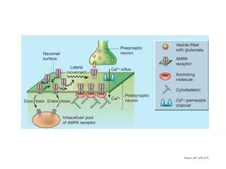
長期給予嗎啡或安非他命對大白鼠或幼鼠腦部興奮性的表現與NMDA受體所產生的影響長期給予嗎啡或安非他命對大白鼠或幼鼠腦部興奮性的表現與NMDA受體所產生的影響 • 第一部份 長期給予母鼠嗎啡對於其幼鼠腦部NMDA receptor 各亞型表現的影響中文摘要 嗎啡成癮的母親所生的小孩常會有著長期神經、精神異常的現象,然而引發的機制尚未明瞭。在過去本實驗室已經證實,長期投與嗎啡的母鼠所生的幼鼠腦中,N-methyl-D-aspartate receptor (NMDA receptor,一種興奮性胺基酸的接受體之一)的密度在出生後第十四天比起控制組有明顯的下降。由此顯示,長期嗎啡成癮的幼鼠腦部神經細胞內的NMDA receptor會有減量調控(down-regulation)的現象。過去有實驗證實,NMDA receptor是由數個亞型所組合而成,包括NR1A, NR2A, NA2B, NR2C, NR2D。因此,長期給予嗎啡可能與腦部NMDA receptor各亞型產生減量調控的變化有關。本研究於是利用西方墨點法來定量幼鼠腦中NMDA receptor各亞型的變化,以求更進一步瞭解幼鼠腦中NMDA receptor表現的亞型專一性。我們採取遵循過去本實驗室的嗎啡給藥模式,並在幼鼠出生後的第七天、十四天、二十八天、六十天將其予以斷頭犧牲,取腦分為大腦皮質、海馬迴等各部位分別製備成非溶解性蛋白質溶液,利用NR1A, NR2A, NA2B之專一性抗體來量化各亞型,並以neuron-specific enolase (NSE)來偵測神經細胞量的多寡以作為內部標準控制。本研究結果發現,在嗎啡組的大腦皮質中,NR1A的表現量在出生後第十四天大約少於對照組40%,NR2A的表現量則是在出生後第七天大約比對照組增加2.8倍,而NR2B的表現量與對照組間無明顯差異。在嗎啡組的海馬迴中,NR1A及NR2B的表現量與對照組間並無明顯差異,但是NR2A的表現量則是在出生後第十四天大約少於對照組34 %。此結果與本實驗室過去利用受體結合試驗觀察NMDA receptor的變化結果相吻合;並且更進一步顯示長期曝露在嗎啡下的幼鼠,其腦中NMDA receptor各亞型表現的減量調控,在大腦皮質中為NR1A,在海馬迴中為NR2A。第二部份 長期給予安非他命對於kainic acid誘發大白鼠癲癇發作 行為變化及腦部NMDA receptor各亞型表現的影響中文摘要 安非他命,一種神經興奮劑,是成癮與濫用的主要藥物之一,對於社會及公共衛生形成重要的衝擊。長期重覆使用安非他命者,會產生非常類似妄想性精神分裂的精神病變,此種安非他命引起之精神病經常在戒除藥物後即消失殆盡,然而有一部分安非他命成癮者,即使停藥了幾年後,當再次投與低劑量的安非他命時,會出現劇烈反應,包括情緒不穩定、精神症或癲癇等的症狀。引發對安非他命此種高度敏感化的機制目前仍未清楚,但大腦興奮性的改變可能是其中一項因素。此外在過去本實驗室已經證實,重覆投與安非他命的大白鼠大腦皮質中,N-methyl-D-aspartate receptor (NMDA receptor,一種興奮性胺基酸的接受體之一) 的密度在安非他命停藥後的第十四及二十八天,比起控制組有明顯的增高。如此顯示,重覆投與安非他命的大白鼠腦部神經細胞的NMDA receptor會有增量調控(up-regulation)的現象。所以我們推測,長期重覆對大白鼠給予安非他命可能會影響腦部NMDA receptor的表現。因此本研究一方面利用kainic acid引發癲癇的行為變化,來探討重覆給與安非他命後,對於大腦興奮性是否有改變的現象。另一方面利用西方墨點法來定量大白鼠腦中的NMDAreceptor各亞型的變化,以求更進一步瞭解大白鼠腦中NMDA receptor表現的亞型專一性。我們採用Sprague-Dawley大白鼠每日給與一次腹腔注射安非他命(5mg/kg) 連續14天,然後分別在安非他命停藥後的第一、十四、二十八天皮下注射一次劑量的kainic acid(12mg/kg),並且觀察3小時內kainic acid誘發抽搐的程度及其引發抽搐的潛伏時間。控制組則每日腹腔注射生理食鹽水。隔日將大白鼠予以斷頭犧牲,取腦分為大腦皮質、海馬迴等各部位分別製備成非溶解性蛋白質溶液,利用NR1A, NR2A, NR2B之專一性抗體來量化各亞型,並以neuron-specific enolase (NSE)來偵測神經細胞量的多寡以作為內部標準控制。 在本研究的大白鼠抽搐行為觀察中發現,在控制組由kainic acid引發產生wet-dog shakes的頻率,於停止生理食鹽水注射後的第一天及第二十八天,比起第十四天有減少的現象。安非他命組則在停藥後的第一天及第二十八天,產生wet-dog shakes的頻率明顯的高於同時期的控制組。安非他命組在停藥後的第十四天,引發產生wet-dog shakes的潛伏時間比控制組慢了31.5 %。安非他命組在停藥後的第一天,由kainic acid引發嚴重抽搐發作的隻數比例比起控制組有明顯的增加,然而在其餘停藥後的天數則沒有明顯變化。在本研究西方墨點法的結果發現,在安非他命組的大腦皮質中,NR1A的表現量在停止給予安非他命後的第一天,明顯的少於控制組約68 %。而NR2A, NR2B, NSE,以及在海馬迴中的NR1A, NR 2A, NR2B, NSE則無此現象發生。 由本研究以上的結果我們推測,長期給予安非他命後會顯著增加對大腦的興奮性。並且KA 注射會選擇性的對安非他命組大腦皮質的NR 1A產生短暫性的減量調控。這種現象有可能是與慢性給與安非他命導致KA引發腦部興奮性的增加有關。
The effect of chronic treatment of morphine or amphetamine on the expression of excitability and N-methyl-D-Aspartate (NMDA) receptor subunits in rat brain • Part 1 :abstract : Long-term neuropsychological abnormality has been documented in child born to morphine addicted mothers, but the mechanism of this phenomenon until now is not clear. Our previous ligand binding study has shown that combined prenatal and postnatal chronic treatment of morphine could alter the ontogenic expression of the NMDA receptor. In that, the density of NMDA receptor of rats born to dam rats received morphine treatment were significantly less than the control rats on postnatal day 14 (PND 14) . It suggested that rats born to chronic morphine addicted dam rats could induce the neuronal NMDA receptor downregulation. To further explore what kinds of NMDA receptor subunits are changed, we used western blotting to examine the expression of the NMDAreceptor subunits, namely, (NR1A, NR2A, NR2B) proteins in the brain of rats born to morphine-treated dam rats. In cortex we found that the NR1A of morphinerats was decreased by 40 % on PND 14 as compare to that of control group. Onthe contrary, the NR2A of morphine rats on PND7 was increased 2.8 folds as compared to that of control rats. But the NR2B of morphine rats was not differentto that of control rats. In hippocampus, the NR1A and NR2B of morphine rats were not different to that of control rats. However, the NR2A of morphine ratswas decreased by 34 % as compared to that of control rats. These results are in consistent with our previous ligand binding assay, suggest that rats born tochronic morphine addicted dam rats induce cerebral NMDA receptor subunits downregulation, in the cortex that is NR1A and in the hippocampus that is NR2A. Part 2:abstract: Addition to the psychstimulant drugs, such as amphetamine (AMPH), is one of the important social and public health problems in Taiwan. Repeated administration of amphetamine frequently develop a drug-induced paranoidpsychosis, and the this phenomenon usually dissipates upon the cessation of drug use. But there are some long-term sequel associated with amphetamine abuse, such as, re-exposure to a relatively low dose of amphetamine will often precipitate a psychotic episode in former amphetamine addicts who have been abstinent for months to years. The mechanism of amphetamine produces hypersensitivity is not clear, however, it may be associated with the change of cerebral excitability. Previous investigations of the mechanism by AMPH induced addiction and long-term neurotoxicity is mainly focusing on dopaminergic neuron, however,recent evidenced have indicated that the N-methyl-D-aspartate receptor (NMDAreceptor), one of the subtype receptor of glutamate receptors, which antagonists significantly attenuated AMPH-induced behavioral sensitization, glutamate release. To explore the amphetamine induced long-term neurotoxicity, we studied the effect of chronic treatment of amphetamine in kainic acid (KA) elicitedseizure activity. Male Sprague-Dawley rats were received daily i.p. injection
of 5 mg/kg amphetamine for 14 days. The animals were received a single dose of kainic acid (12 mg/kg) s.c. injection 1, 14 and 28 days after the last injection of amphetamine. We observed the behavior change during the first 3h after. Control rats were received daily injection of normal-saline before injectionof kainic acid. Furthermore, we used western-blotting to quantify the densityof each NMDA receptor subunits, namely, the NR1A, NR2A, NR2B in the crude membrane of cortex and hippocampus tissues prepared from rats received single dose of KA after they had received 14 day injection of AMPH (AMPH group) or saline (control) group. The results of kainic acid-induced seizure showed that in control group rats, the wet-dog shake frequency was significant lower at 1 and 28 days than that of 14 days after the last injection of normal-saline. Inamphetamine-treated group, the wet-dog shake frequency was significant higherthan control group at 1 and 28 days after the last injection of amphetamine. The latency of wet-dog shakes onset was later than control group about 31.5 % at 14 days after the last injection of amphetamine. The percentage of rats hadmotor clonus of amphetamine group is significant higher than that of control group rats at 1 day after the last injection of amphetamine. The results of western boltting showed a significant decrease in the density of NR1A in the cortex of AMPH group one day after last injection of AMPH. However, no significantdifference in the expression of this subunit in other time points or hippocampus between control and AMPH group. Furthermore, there is no significant change in the expression of NR2A and NR2B of AMPH group. These results suggestedthat chronic administration of amphetamine lead to a transiently enhancing the kainic acid-induced neuroexcitation, and demonstrated that chronic treatmentof AMPH with single injection of KA may produce a transient down-regulation of one NMDA receptor subunit. This receptor plasticity may be a consequence ofchronic treatment of AMPH which result into an enhanced KA-induced neurotoxicity.