Download
1 / 2
20 likes | 147 Views
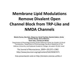
(1) 安非他命對 NMDA 在大白鼠大腦皮質神經細胞上所引起 45Ca2+ 蓄積之作用 (2) 在大白鼠大腦皮質神經細胞培養中發生的細胞凋零的特徵.

E N D
(1) 安非他命對NMDA在大白鼠大腦皮質神經細胞上所引起45Ca2+蓄積之作用 (2) 在大白鼠大腦皮質神經細胞培養中發生的細胞凋零的特徵 1)安非他命是一種精神刺激劑,有關安非他命的中樞神經作用的研究過去大都著重在與多巴胺神經傳導系統的關係,然而最近有許多證據顯示安非他命所造成的行為敏感化作用 (behavior sensitization) 能被N-methyl-D-aspartate receptor (NMDA) 的拮抗劑所阻斷, NMDA受體的活化參與了安非他命的中樞神經的作用。過去本實驗室利用[3H]TCP受體結合的方法及45Ca2+ accumulation的實驗,觀察安非他命對NMDA受體的作用,結果顯示安非他命可以直接抑制NMDA受體的活化。在本實驗中我們將再次確認安非他命可以抑制NMDA所引起的45Ca2+ accumulation,並且比較右旋 (d-amphetamine)、左旋安非他命 (l-amphetamine)及甲基安非他命 (Methamphetamine)對NMDA所引起的45Ca++ accumulation是否具有抑制作用及其個別的強度。結果顯示,安非他命是NMDA的拮抗劑且安非他命對NMDA引起的45Ca2+ accumulation有兩階段的抑制作用。而不同結構的安非他命對NMDA引起的45Ca2+ accumulation的抑制作用強度是差不多的。(2) Apoptosis (細胞凋亡)又稱為Programmed cell death (計劃性細胞死亡),顧名思義,它是由細胞自行調控死亡,是一種選擇性的主動死亡方式。本實驗利用大白鼠大腦皮質神經細胞的培養觀察apoptosis的現象,我們發現隨著培養天數的增加,神經細胞的密度漸漸降低而且有許多細胞呈現apoptosis的特徵包括神經細胞縮小、核質濃縮、及產生許多apoptotic bodies等。因此我們判定這種神經細胞培養有自生性的apoptosis,根據此種觀察,我們想要利用其它重要方法來確定此種判斷。我們除了利用倒立式相位差顯微鏡直接觀察並照下神經細胞的外型之外,並且以LDH response測量細胞毒性,及利用TUNEL assay 偵測apoptosis的細胞,以及利用RT-PCR的方法偵測一些mRNA的表現量,包括bax、c-fos、c-jun等。此外,我們並分別在神經細胞離體外培養第6、12、18天時投與NMDA、MK-801、morphine、naloxone等藥物觀察它們對神經細胞的影響,瞭解他們是否會促進或抑制神經細胞的apoptosis。結果發現大白鼠大腦皮質初代神經細胞培養 (rat primary neuronal cell culture)中有自發性的apoptosis,可作為一種研究apoptosis的模式。而且直接以顯微鏡及以TUNEL assay觀察都發現:apoptosis細胞的數目會隨著培養天數增加而上升。再偵測培養1到22天的細胞的mRNA,以ß-actin作為internal control,發現c-fos mRNA的量在第20天有明顯的上升,但是NSE、NF-M、bax、c-jun mRNA在不同天數則沒有明顯差異。在第6天的culture上不論NMDA、MK-801、morphine、naloxone對細胞內LDH釋放的量都沒有明顯的作用,細胞型態在不同時間也沒有明顯變化,但是投與NMDA與MK-801會降低c-fos mRNA的量。在第12、18的細胞投與NMDA與MK-801則會降低NSE和NF-M mRNA的量。在第12天、18天的culture上NMDA組的LDH值比control組高,在倒立式相位差顯微鏡下觀察到NMDA組細胞外型呈現明顯的necrosis,到24小時細胞已明顯的死亡(Figure 27,29),此時以TUNEL assay偵測apoptosis的細胞,發現neuron的數目較少,而且存活下來的細胞也幾乎100%發生apoptosis。可見得NMDA會同時造成neuron的necrosis與apoptosis。
(1) The Effect of Amphetamine on the N-Methyl-D-Aspartate Induced Intracellular 45Ca2+ Accumulation.(2) The Characterization of the Apoptosis in Rat Primary Cortical Cell Culture. • (1) Recent investigations have indicated that amphetamine, a psychostimulator, produces its central effect by activation of the NMDA (N-methyl-D-aspartate) receptor, a subtype receptor of the glutamate receptor. Our previous study using [3H]TCP receptor ligand binding to assess this receptor/channel complex had shown that amphetamine could directly inhibit the NMDA-coupled ion channel by acting at multiple sites on the receptor. In the present study, we examined this specific action of amphetamine and compare the effect of d-amphetamine、l-amphetamine、Methamphetamine on the NMDA receptor-mediated 45Ca2+ accumulation. • This study confirm that amphetamine could direct inhibit the function of NMDA receptor, and the d-amphetamine、l-amphetamine、Methamphetamine have similar effect on the NMDA receptor-mediated 45Ca2+ accumulation. • (2) Apoptosis (also termed programmed cell death, PCD) is a mode of cell death in which the cell plays an active role in its demise. In the present study, we investigate the characteristic of apoptosis in the primary cortical neuronal culture. We find that the cell in our rat primary cortical cell culture undergoing apoptosis by the examination of microscope. We confirm the presence of apoptosis in this culture by several biochemical methods. Including taking picture of neuronal culture under microscope, examine the apoptotic cells by TUNEL assay and immunocytochemistry, and quantify the mRNA level of bax, c-fos, c-jun by RT-PCR. Besides, we also examine the LDH response of culture cells. In addition, we also detect the effect of NMDA (N-methyl-D-aspartate) (100M), MK-801 (10M), morphine (10M), and naloxone (10M) on the apoptosis of this culture in DIV6, 12, 18 culture. • We find that our rat primary cortical cell culture undergoing spontaneous apoptosis expressed as increase in the LDH level, increase in the number of TUNEL positive neuron in a two-phase manner. This apoptotic phenomenon is accompanied with increase gene expression of c-fos, and likely the expression of bax gene. Addition of NMDA to culture also induces significant increase in LDH response accompanies with apoptosis. But MK-801, morphine, or naloxone has no effect on LDH response and apoptosis. However, NMDA or MK-801 decrease c-fos on DIV 6 culture, but increase c-fos on DIV 18 culture. Morphine and naloxone didn’t have any effect on the c-fos gene expression. NMDA increase apoptotic neurons on DIV 12 and 18 culture, but it has no effect on DIV 6 culture. This study confirm that NMDA increase apoptosis in this culture, but MK-801, morphine, or naloxone didn’t have any effect on the apoptosis in this culture.