Download
1 / 56
560 likes | 560 Views
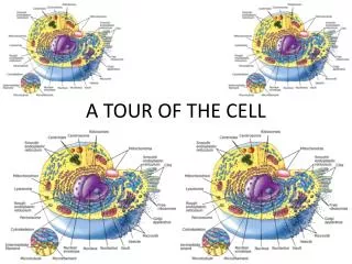
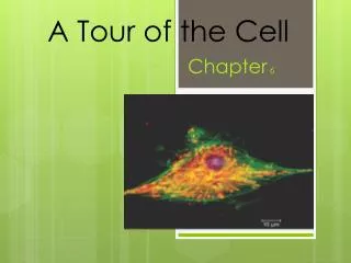
A Tour of the Cell. Chapter 7. Figure 7.1 The size range of cells. Figure 7.3 Cell Fractionation. Biochemical approach in cell biology. Goal is to take cells apart, so that organelles can be separated and their functions can be studied. PROCESS: homogenization – disruption of cells

E N D
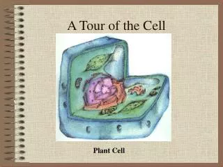
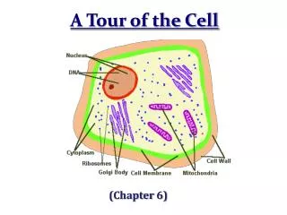
A Tour of the Cell Chapter 7
Figure 7.3 Cell Fractionation • Biochemical approach in cell biology. Goal is to take cells apart, so that organelles can be separated and their functions can be studied. • PROCESS: • homogenization – disruption of cells • low speed in centrifuge – separates into pellet and supernatant • increase speed with each step, collect smaller and smaller organelles
Cells – The Basic Unit of Life • CELLS ARE THE BASIC UNIT OF STRUCTURE AND FUNCTION FOR LIVING THINGS. • ALL LIVING THINGS ARE MADE OF CELLS. • CELLS ARISE FROM PRE-EXISTING CELLS. • Collectively, these are known as the Cell Theory!
Prokaryotes vs. Eukaryotes • Prokaryotes: • NO NUCLEUS, but do have nucleoid region with DNA present • Small and Simple – few organelles • Have cell membranes and cytoplasm • Ex. Bacteria • Eukaryotes: • Contain nuclei • Contains organelles that perform specialized functions • Unicellular or multicellular • Ex. Plant and animal cells
Figure 7.4 A Prokaryotic Cell Lacking a true nucleus and the other membrane-enclosed organelles of the eukaryotic cell, the prokaryotic cell is much simpler in structure. Only organisms of the domain BACTERIA and ARCHAEA have prokaryotic cells.
Basic Cell Parts: Cell Membranehttp://bcs.whfreeman.com/thelifewire/content/chp04/0402001.html • Cell membrane – • provides barrier between internal and external environment of cell • is semi-permeable (some things can go in, some cannot; some things can exit, some never can) • made up of phospholipid bilayer with proteins embedded that allow for needed passage of large molecules
Function of Cell Membrane • Major job of cell membrane is to maintain the cell’s environment – establish homeostasis!
Problem: Surface Area to Volume Ratio • Surface area acts as limiting factor in size of cell because is a two dimensional unit. • Volume is three dimensional, so increases more quickly than the surface area can accommodate. • Larger organisms do not generally have LARGER CELLS, simply MORE CELLS!
Surface Area to Volume Ratio Problem • Metabolic requirements impose upper limits on cell size. • As an object increases in size, its volume grows proportionately more than surface area (area2 and volume3)… • So…the smaller the object, the greater its ratio of surface area to volume. • As an object increases in size its volume increases as the cube of its linear dimensions while surface area increases as the square. • As these cubes illustrate the surface area to volume ratio of a small object is larger than that of a large object of similar shape. This ratio limits how large cells can be.
Figure 8.6 The detailed structure of an animal cell’s plasma membrane, in cross section
Fluid Mosaic Model • Cell membrane and embedded proteins are not locked into position – they flow against one another as the cytoplasm and the external liquid environment dictate.
Other Components of the Cell Membrane • Cholesterol – helps to stabilize the phospholipids
Proteins in Cell Membrane • Channel proteins – act as passageways through the phospholipid bilayer for large things: • These are integral proteins – penetrate the hydrophobic core of the lipid bilayer. • Receptor proteins – receive info about the environment outside the cell and transmit it into the cell – no physical molecule passes through this, just INFO: • may be integral or peripheral proteins – not embedded in the bilayer, can be extensions of integral proteins • Marker Proteins – identify the type of cell using carbohydrate chains (glycoproteins) • may also have glycolipids that are not attached to proteins
Cytoplasm • CYTOPLASM includes the entire region between the nucleus and the cell membrane! • The semi-fluid substance that fills this area is called CYTOSOL, and this is what the organelles are suspended in.
Cell Wall • Found in plant cells (another barrier in ADDITION to the cell membrane). • Protects the cell. • Gives support to cell. • Made of polysaccharide called cellulose. • Is very porous and allows molecules to pass through, but is NOT SELECTIVELY PERMEABLE!!!
Organelles • Control: *Nucleus (plant and animal) *Centrosome (plant and animal) • Assembly, Transport, and Storage: *Endoplasmic reticulum (plant and animal) *Ribosomes (plant and animal) *Golgi apparatus (plant and animal) *Vacuoles (plant -1 large, and animal - many) *Lysosomes (animal) *Leucoplasts (plant only) • Energy transformations: *Chloroplasts and Chromoplasts (plant only) *Mitochondria (plant and animal)
Some Useful Animation Tutorials • http://bcs.whfreeman.com/thelifewire/content/chp04/0402001.html • www.cellsalive.com
Nucleus • Contains most of eukaryotic cell’s genetic library • Largest organelle • Enclosed by nuclear envelope or membrane, which is a doublemembrane – each of which is a lipid bilayer!!! • Nuclear envelope has pores in it • Nuclear lamina lines nuclear side of envelope – a net-like array of protein filaments that maintain nuclear shape. • May also be a nuclear matrix that extends into the entire nucleus • Contains inactive DNA – chromatin • When gets ready to divide, chromatin condenses into chromosomes • Directs protein synthesis by synthesizing mRNA and sending to ribosomes in the cytoplasm • DNA mRNA protein (transcription and translation)
Nucleolus • Prominent structure in non-dividing nucleus • Components of ribosomes are synthesized here (ribosomal RNA and ribosomal subunits)
Ribosomes • Sites of PROTEIN SYNTHESIS • Are made of rRNA and protein • Made of two subunits – • one acts as attachment point for beginning of translation, • other acts as “cap” to prevent mRNA from moving until tRNA has brought in appropriate amino acid • Cells with high rates of protein synthesis have MANY ribosomes (human pancreas cell has MILLIONS of ribosomes) • There are “free” ribosomes in cytosol that make proteins for the cell in which they reside • Ribosomes that are attached to endoplasmic reticulum (bound) are making proteins for packaging and export
Endomembrane System • Many of the different membranes of the eukaryotic cell are part of an ENDOMEMBRANE SYSTEM. • Membranes in cell are not identical in structure or function (modifications are present according to job) • Includes: • nuclear envelope • endoplasmic riticulum • Golgi apparatus, • Lysosomes • Vacuoles • plasma membrane
Figure 7.16 Review: relationships among organelles of the endomembrane system
Endoplasmic Reticulum • A membrous system of channels and flattened sacs that traverse the cytoplasm • 2 varieties: • Rough ER: the site of protein synthesis resulting from the attached ribosomes • Smooth ER: assists in the synthesis of steroid hormones and other lipids • Also connects rough ER to the Golgi apparatus and carries out various detoxification processes in liver • Smooth and rough E.R. are actually connected, not distinct, separate sections
Golgi Apparatus • Packages substances produced in the rough ER and secretes them to other cell parts or to the cell surface for export. • Has cis (entrance) side and trans (exit) side • Golgi will modify products as needed – gives more variety by removing some monomers and substituting others • Knows what to do by using molecular identification tags (like phosphate groups); even adds molecules on their membranes that may recognize “docking sites” on organelle surfaces or on the cell membranes of other cells
Lysosomes • Membrane-bounded sac of hydrolytic enzymes – are the principle site of intracellular digestion • Different lysosomes break down each of the major classes of macromolecules – proteins, polysaccharides, fats, nucleic acids • Work best at pH of 5 • Using active transport to maintain this – pumps hydrogen ions from cytosol into itself • Used in autophagy – recycle the cell’s own organic material for use • Can also be used in programmed destruction of cells by lysosomal enzymes – ex. Tadpole loses tail • Related diseases – Pompe’s and Tay-Sachs (pg. 122)
Figure 7.14 The formation and functions of lysosomes (Layer 1)
Figure 7.14 The formation and functions of lysosomes (Layer 2)
Figure 7.14 The formation and functions of lysosomes (Layer 3)
Animated Tutorial – Endosymbiotic Theory • http://www.sumanasinc.com/webcontent/animations/content/organelles.html
Peroxisomes • Job is to generate and degrade hydrogen peroxide—contain enzymes that transfer hydrogen from various substrates and make H2O2 as a by-product • Also detoxify alcohol in liver cells • H2O2 is toxic, but peroxisomes contain enzymes that convert it to water.
Cytoskeleton • Network of fibers extending into cytoplasm of cell • Provides structural support, and aids in cell motility and cell regulation • Made up of microtubules (thickest), microfilaments (thinnest), and intermediate filaments (see page 127)
Centrosome and Centrioles • Microtubules often grow out of centrosome (central area of cell) • Within the centrosome are centrioles, each composed of nine sets of triplet microtubules arranged in a ring; CENTRIOLES aid in chromosome separation
Cilia and Flagella • The movement of these two locomotor appendages is controlled by microtubules • Cilia are short projections, flagella are much longer • Movement may not be for entire organism; may be part of a larger unit – ex. Cilia lining windpipe propel foreign substances out… • See page 129 for diagrams
Figure 7.23 A comparison of the beating of flagella and cilia
Figure 7.24 Ultrastructure of a eukaryotic flagellum or cilium
Dynein • Large protein that makes up the motor molecules that extend from each microtubule doublet to the next • Responsible for the bending and movement of cilia and flagella • See page 130